Manganese »
PDB 7sd0-7u7z »
7tu2 »
Manganese in PDB 7tu2: Structure of the L. Blandensis Dgtpase R37A Mutant Bound to Mn
Protein crystallography data
The structure of Structure of the L. Blandensis Dgtpase R37A Mutant Bound to Mn, PDB code: 7tu2
was solved by
A.P.Sikkema,
B.P.Klemm,
J.C.Horng,
T.M.T.Hall,
with X-Ray Crystallography technique. A brief refinement statistics is given in the table below:
| Resolution Low / High (Å) | 36.22 / 2.13 |
| Space group | P 41 21 2 |
| Cell size a, b, c (Å), α, β, γ (°) | 179.934, 179.934, 110.941, 90, 90, 90 |
| R / Rfree (%) | 20.4 / 22.6 |
Manganese Binding Sites:
The binding sites of Manganese atom in the Structure of the L. Blandensis Dgtpase R37A Mutant Bound to Mn
(pdb code 7tu2). This binding sites where shown within
5.0 Angstroms radius around Manganese atom.
In total 3 binding sites of Manganese where determined in the Structure of the L. Blandensis Dgtpase R37A Mutant Bound to Mn, PDB code: 7tu2:
Jump to Manganese binding site number: 1; 2; 3;
In total 3 binding sites of Manganese where determined in the Structure of the L. Blandensis Dgtpase R37A Mutant Bound to Mn, PDB code: 7tu2:
Jump to Manganese binding site number: 1; 2; 3;
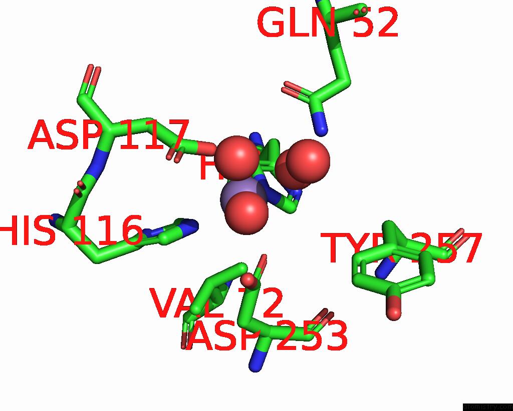
Manganese binding site 1 out of 3 in 7tu2
Go back to
Manganese binding site 1 out
of 3 in the Structure of the L. Blandensis Dgtpase R37A Mutant Bound to Mn

Mono view
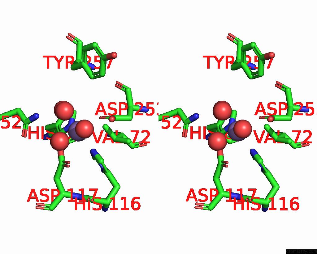
Stereo pair view
Mono view
Stereo pair view
A full contact list of Manganese with other atoms in the Mn binding
site number 1 of Structure of the L. Blandensis Dgtpase R37A Mutant Bound to Mn within 5.0Å range:
|
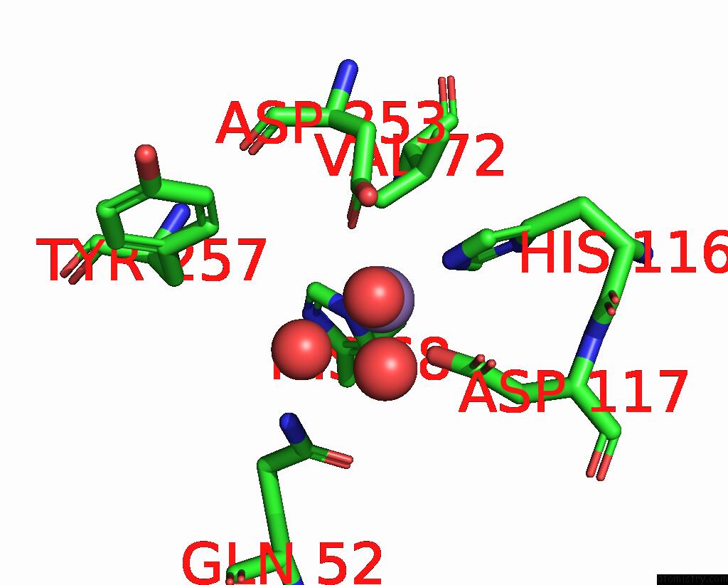
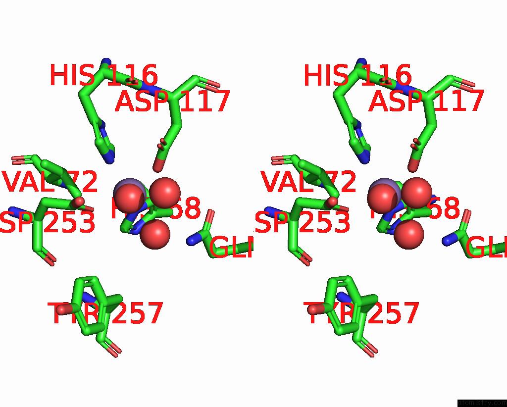
Manganese binding site 2 out of 3 in 7tu2
Go back to
Manganese binding site 2 out
of 3 in the Structure of the L. Blandensis Dgtpase R37A Mutant Bound to Mn
Mono view

Stereo pair view
Mono view
Stereo pair view
A full contact list of Manganese with other atoms in the Mn binding
site number 2 of Structure of the L. Blandensis Dgtpase R37A Mutant Bound to Mn within 5.0Å range:
|
Manganese binding site 3 out of 3 in 7tu2
Go back to
Manganese binding site 3 out
of 3 in the Structure of the L. Blandensis Dgtpase R37A Mutant Bound to Mn
Mono view
Stereo pair view
Mono view
Stereo pair view
A full contact list of Manganese with other atoms in the Mn binding
site number 3 of Structure of the L. Blandensis Dgtpase R37A Mutant Bound to Mn within 5.0Å range:
|
Reference:
B.P.Klemm,
A.P.Sikkema,
A.L.Hsu,
J.C.Horng,
T.M.T.Hall,
M.J.Borgnia,
R.M.Schaaper.
High-Resolution Structures of the SAMHD1 Dgtpase Homolog From Leeuwenhoekiella Blandensis Reveal A Novel Mechanism of Allosteric Activation By Datp. J.Biol.Chem. V. 298 02073 2022.
ISSN: ESSN 1083-351X
PubMed: 35643313
DOI: 10.1016/J.JBC.2022.102073
Page generated: Sun Oct 6 10:37:02 2024
ISSN: ESSN 1083-351X
PubMed: 35643313
DOI: 10.1016/J.JBC.2022.102073
Last articles
Zn in 9MJ5Zn in 9HNW
Zn in 9G0L
Zn in 9FNE
Zn in 9DZN
Zn in 9E0I
Zn in 9D32
Zn in 9DAK
Zn in 8ZXC
Zn in 8ZUF