Manganese »
PDB 7sd0-7u7z »
7tl8 »
Manganese in PDB 7tl8: 1.95A Resolution Structure of Independent Phosphoglycerate Mutase From S. Aureus in Complex with A Macrocyclic Peptide Inhibitor (Sa-D3)
Enzymatic activity of 1.95A Resolution Structure of Independent Phosphoglycerate Mutase From S. Aureus in Complex with A Macrocyclic Peptide Inhibitor (Sa-D3)
All present enzymatic activity of 1.95A Resolution Structure of Independent Phosphoglycerate Mutase From S. Aureus in Complex with A Macrocyclic Peptide Inhibitor (Sa-D3):
5.4.2.12;
5.4.2.12;
Protein crystallography data
The structure of 1.95A Resolution Structure of Independent Phosphoglycerate Mutase From S. Aureus in Complex with A Macrocyclic Peptide Inhibitor (Sa-D3), PDB code: 7tl8
was solved by
L.Liu,
S.Lovell,
K.P.Battaile,
P.Dranchak,
B.Queme,
M.Aitha,
R.H.P.Vanneer,
H.Kimura,
T.Katho,
H.Suga,
J.Inglese,
with X-Ray Crystallography technique. A brief refinement statistics is given in the table below:
| Resolution Low / High (Å) | 46.11 / 1.95 |
| Space group | P 1 21 1 |
| Cell size a, b, c (Å), α, β, γ (°) | 46.898, 74.172, 75.017, 90, 100.54, 90 |
| R / Rfree (%) | 19.4 / 25.5 |
Manganese Binding Sites:
The binding sites of Manganese atom in the 1.95A Resolution Structure of Independent Phosphoglycerate Mutase From S. Aureus in Complex with A Macrocyclic Peptide Inhibitor (Sa-D3)
(pdb code 7tl8). This binding sites where shown within
5.0 Angstroms radius around Manganese atom.
In total 2 binding sites of Manganese where determined in the 1.95A Resolution Structure of Independent Phosphoglycerate Mutase From S. Aureus in Complex with A Macrocyclic Peptide Inhibitor (Sa-D3), PDB code: 7tl8:
Jump to Manganese binding site number: 1; 2;
In total 2 binding sites of Manganese where determined in the 1.95A Resolution Structure of Independent Phosphoglycerate Mutase From S. Aureus in Complex with A Macrocyclic Peptide Inhibitor (Sa-D3), PDB code: 7tl8:
Jump to Manganese binding site number: 1; 2;
Manganese binding site 1 out of 2 in 7tl8
Go back to
Manganese binding site 1 out
of 2 in the 1.95A Resolution Structure of Independent Phosphoglycerate Mutase From S. Aureus in Complex with A Macrocyclic Peptide Inhibitor (Sa-D3)
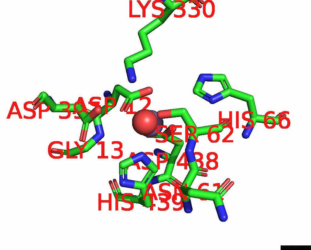
Mono view
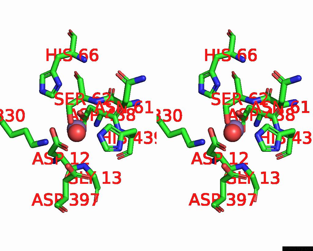
Stereo pair view
Mono view
Stereo pair view
A full contact list of Manganese with other atoms in the Mn binding
site number 1 of 1.95A Resolution Structure of Independent Phosphoglycerate Mutase From S. Aureus in Complex with A Macrocyclic Peptide Inhibitor (Sa-D3) within 5.0Å range:
|
Manganese binding site 2 out of 2 in 7tl8
Go back to
Manganese binding site 2 out
of 2 in the 1.95A Resolution Structure of Independent Phosphoglycerate Mutase From S. Aureus in Complex with A Macrocyclic Peptide Inhibitor (Sa-D3)
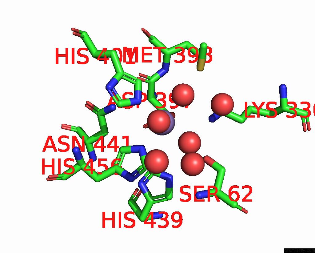
Mono view
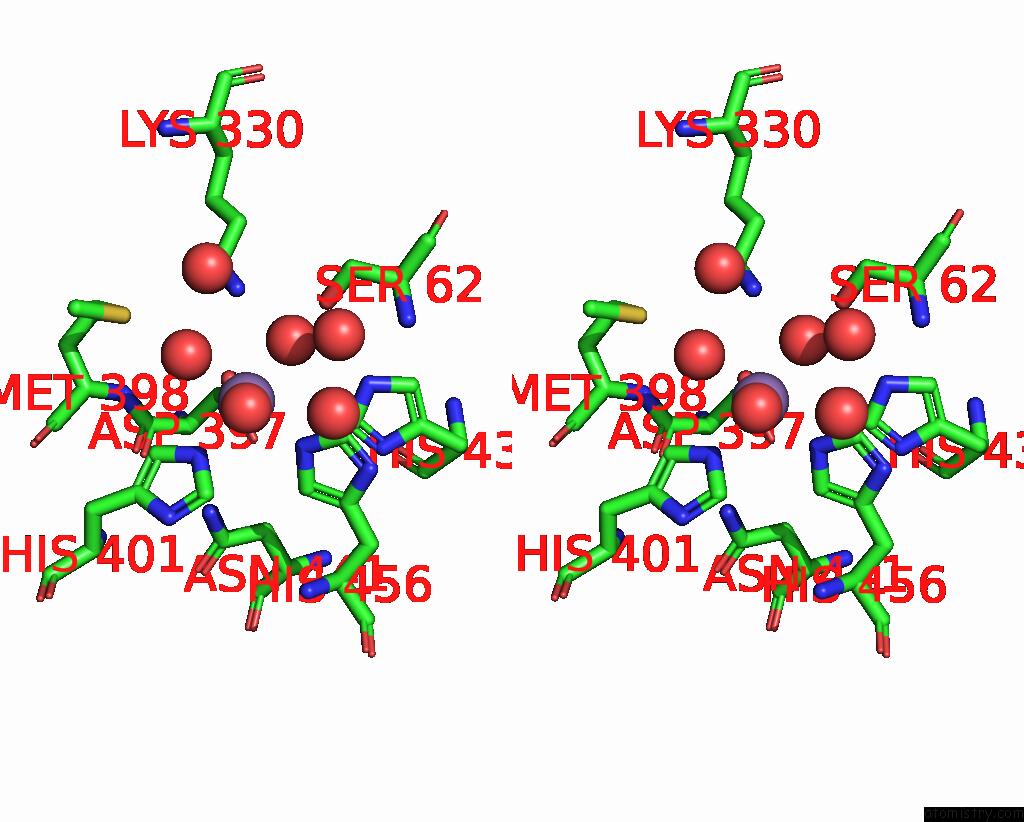
Stereo pair view
Mono view
Stereo pair view
A full contact list of Manganese with other atoms in the Mn binding
site number 2 of 1.95A Resolution Structure of Independent Phosphoglycerate Mutase From S. Aureus in Complex with A Macrocyclic Peptide Inhibitor (Sa-D3) within 5.0Å range:
|
Reference:
R.H.P.Van Neer,
P.K.Dranchak,
L.Liu,
M.Aitha,
B.Queme,
H.Kimura,
T.Katoh,
K.P.Battaile,
S.Lovell,
J.Inglese,
H.Suga.
Serum-Stable and Selective Backbone-N-Methylated Cyclic Peptides That Inhibit Prokaryotic Glycolytic Mutases. Acs Chem.Biol. V. 17 2284 2022.
ISSN: ESSN 1554-8937
PubMed: 35904259
DOI: 10.1021/ACSCHEMBIO.2C00403
Page generated: Sun Oct 6 10:36:29 2024
ISSN: ESSN 1554-8937
PubMed: 35904259
DOI: 10.1021/ACSCHEMBIO.2C00403
Last articles
Zn in 9J0NZn in 9J0O
Zn in 9J0P
Zn in 9FJX
Zn in 9EKB
Zn in 9C0F
Zn in 9CAH
Zn in 9CH0
Zn in 9CH3
Zn in 9CH1