Manganese »
PDB 7sd0-7u7z »
7svs »
Manganese in PDB 7svs: Crystal Structure Analysis of the G73A Mutant of Superoxide Dismutase From Trichoderma Reesei
Enzymatic activity of Crystal Structure Analysis of the G73A Mutant of Superoxide Dismutase From Trichoderma Reesei
All present enzymatic activity of Crystal Structure Analysis of the G73A Mutant of Superoxide Dismutase From Trichoderma Reesei:
1.15.1.1;
1.15.1.1;
Protein crystallography data
The structure of Crystal Structure Analysis of the G73A Mutant of Superoxide Dismutase From Trichoderma Reesei, PDB code: 7svs
was solved by
E.Mendoza Rengifo,
J.R.Ferreira Jr.,
C.R.Garratt,
with X-Ray Crystallography technique. A brief refinement statistics is given in the table below:
| Resolution Low / High (Å) | 72.14 / 2.80 |
| Space group | P 21 21 21 |
| Cell size a, b, c (Å), α, β, γ (°) | 95.17, 110.62, 180.93, 90, 90, 90 |
| R / Rfree (%) | 19 / 25.7 |
Manganese Binding Sites:
The binding sites of Manganese atom in the Crystal Structure Analysis of the G73A Mutant of Superoxide Dismutase From Trichoderma Reesei
(pdb code 7svs). This binding sites where shown within
5.0 Angstroms radius around Manganese atom.
In total 8 binding sites of Manganese where determined in the Crystal Structure Analysis of the G73A Mutant of Superoxide Dismutase From Trichoderma Reesei, PDB code: 7svs:
Jump to Manganese binding site number: 1; 2; 3; 4; 5; 6; 7; 8;
In total 8 binding sites of Manganese where determined in the Crystal Structure Analysis of the G73A Mutant of Superoxide Dismutase From Trichoderma Reesei, PDB code: 7svs:
Jump to Manganese binding site number: 1; 2; 3; 4; 5; 6; 7; 8;
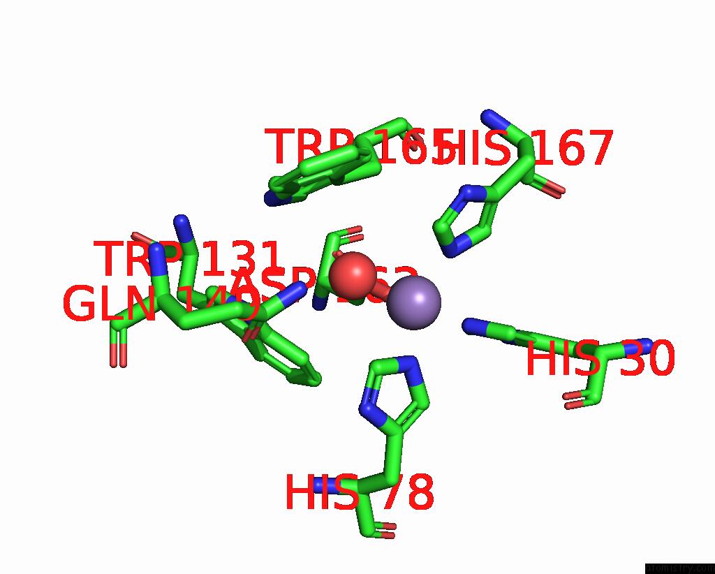
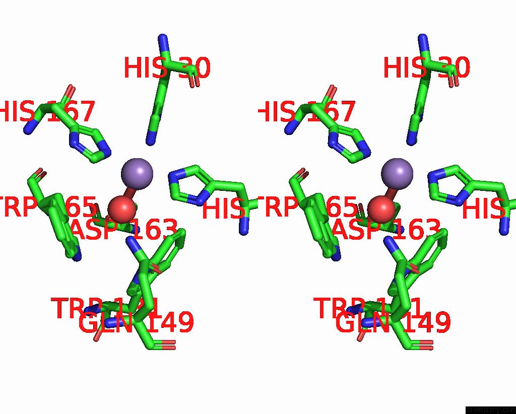
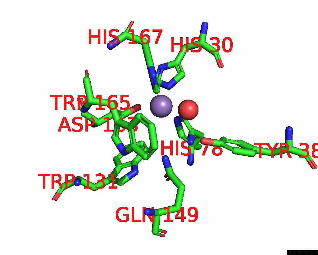
Manganese binding site 1 out of 8 in 7svs
Go back to
Manganese binding site 1 out
of 8 in the Crystal Structure Analysis of the G73A Mutant of Superoxide Dismutase From Trichoderma Reesei
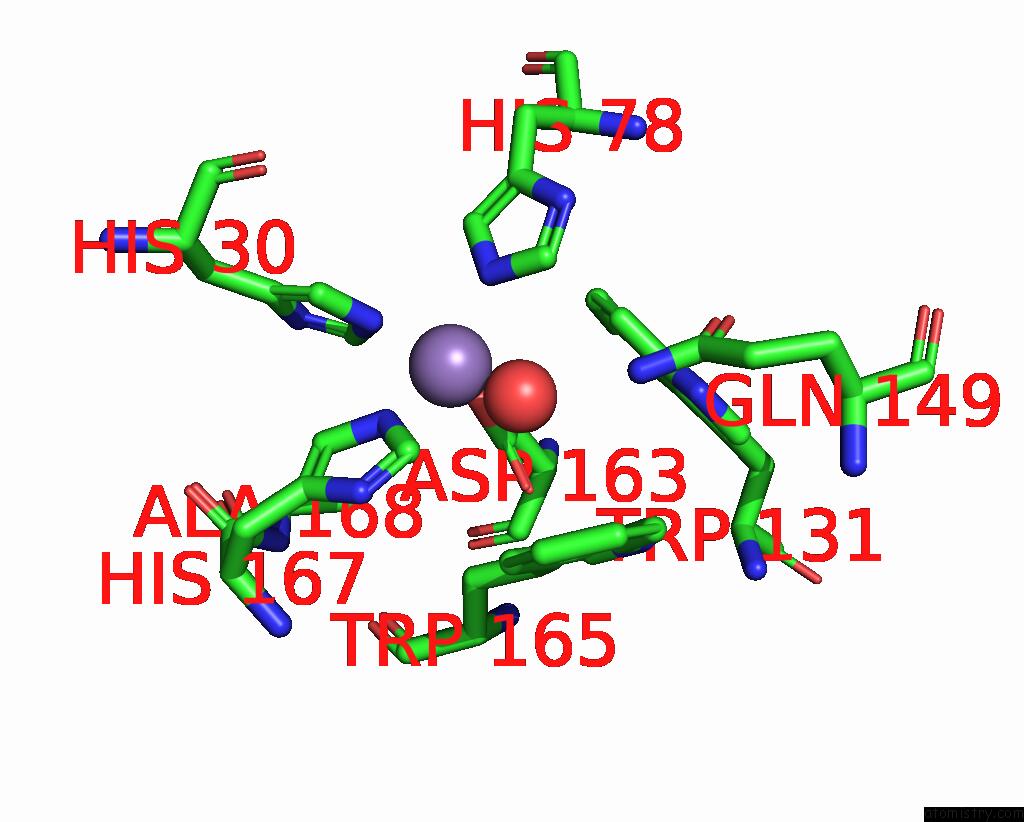
Mono view
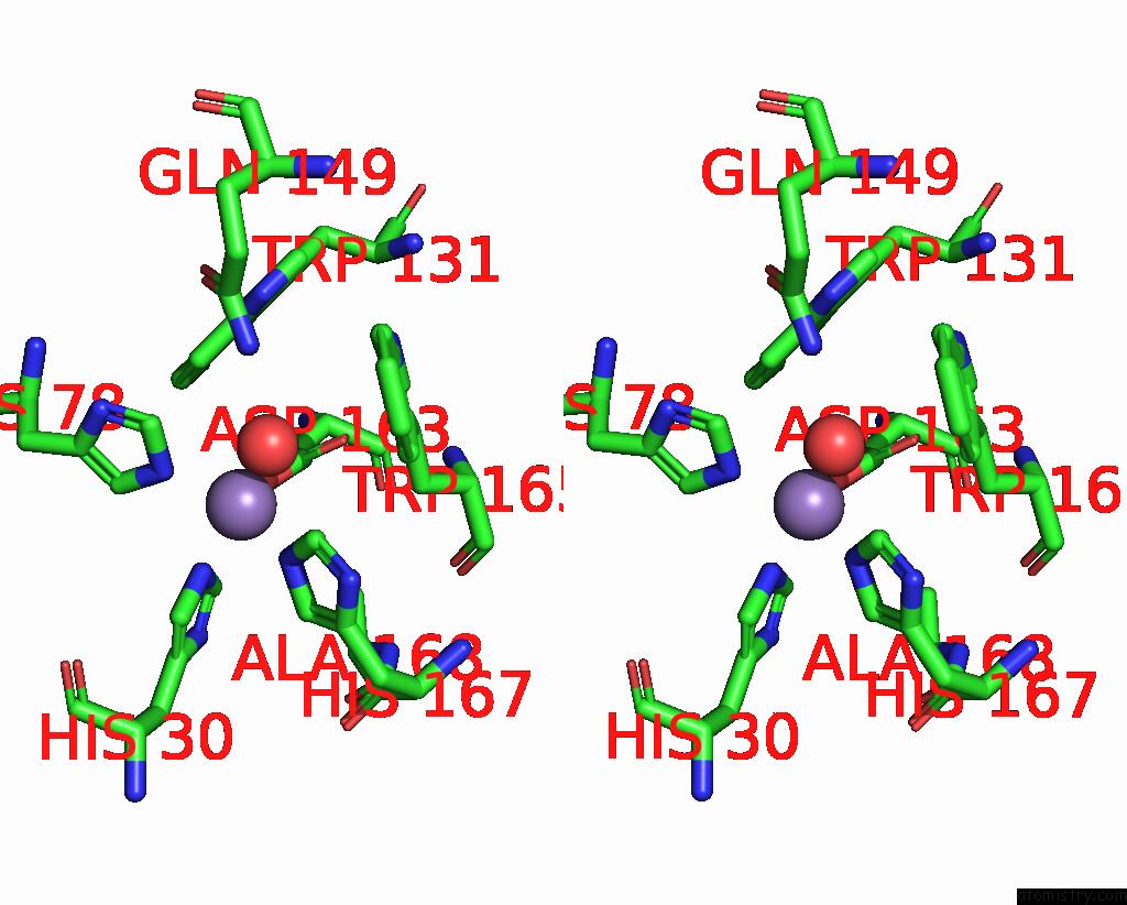
Stereo pair view
Mono view
Stereo pair view
A full contact list of Manganese with other atoms in the Mn binding
site number 1 of Crystal Structure Analysis of the G73A Mutant of Superoxide Dismutase From Trichoderma Reesei within 5.0Å range:
|
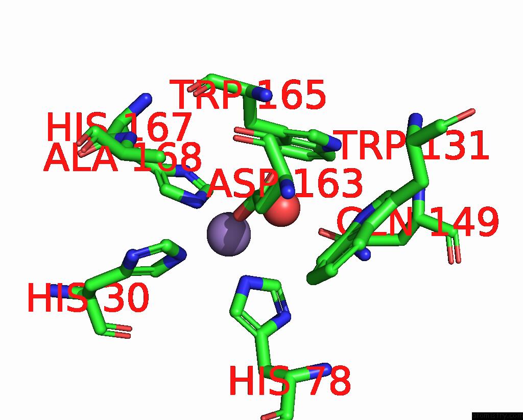
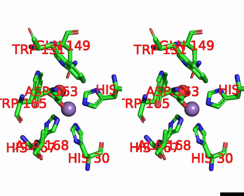
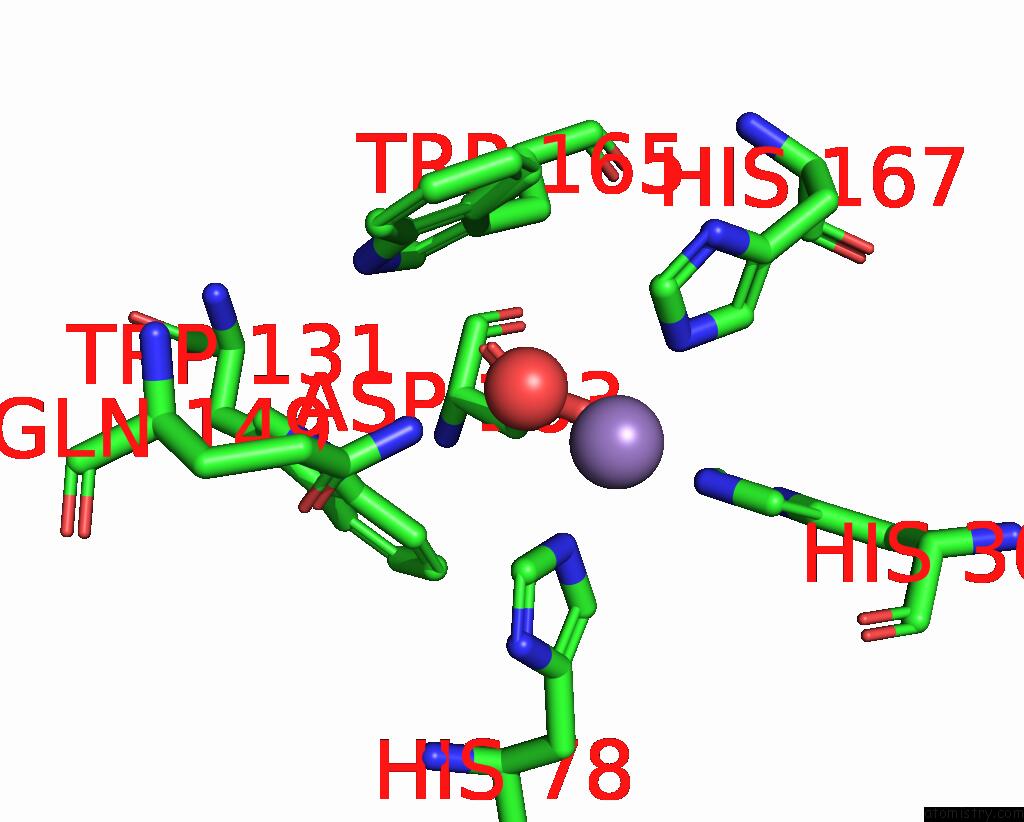
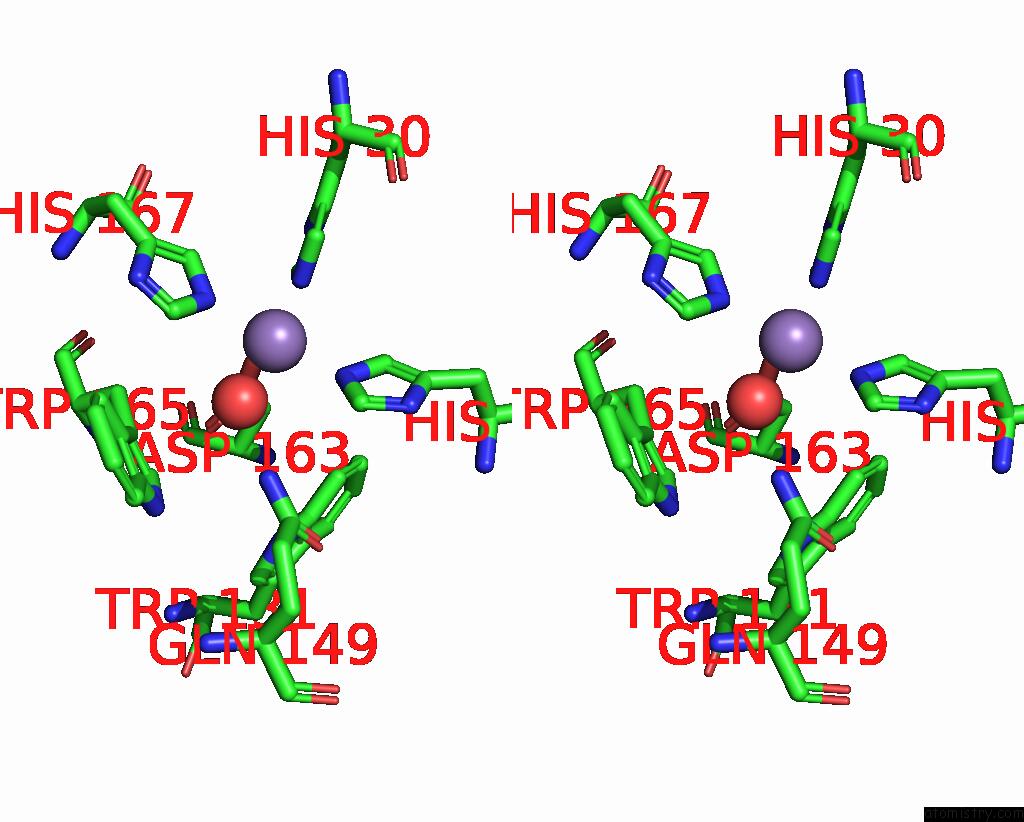
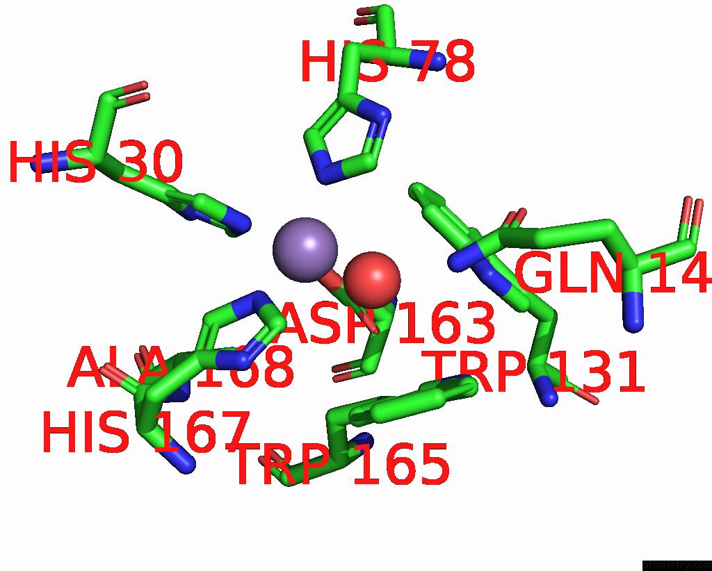
Manganese binding site 2 out of 8 in 7svs
Go back to
Manganese binding site 2 out
of 8 in the Crystal Structure Analysis of the G73A Mutant of Superoxide Dismutase From Trichoderma Reesei

Mono view

Stereo pair view
Mono view
Stereo pair view
A full contact list of Manganese with other atoms in the Mn binding
site number 2 of Crystal Structure Analysis of the G73A Mutant of Superoxide Dismutase From Trichoderma Reesei within 5.0Å range:
|
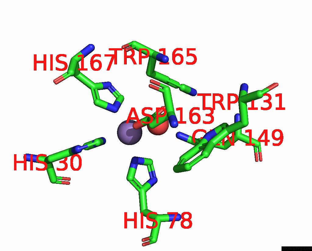
Manganese binding site 3 out of 8 in 7svs
Go back to
Manganese binding site 3 out
of 8 in the Crystal Structure Analysis of the G73A Mutant of Superoxide Dismutase From Trichoderma Reesei
Mono view
Stereo pair view
Mono view
Stereo pair view
A full contact list of Manganese with other atoms in the Mn binding
site number 3 of Crystal Structure Analysis of the G73A Mutant of Superoxide Dismutase From Trichoderma Reesei within 5.0Å range:
|
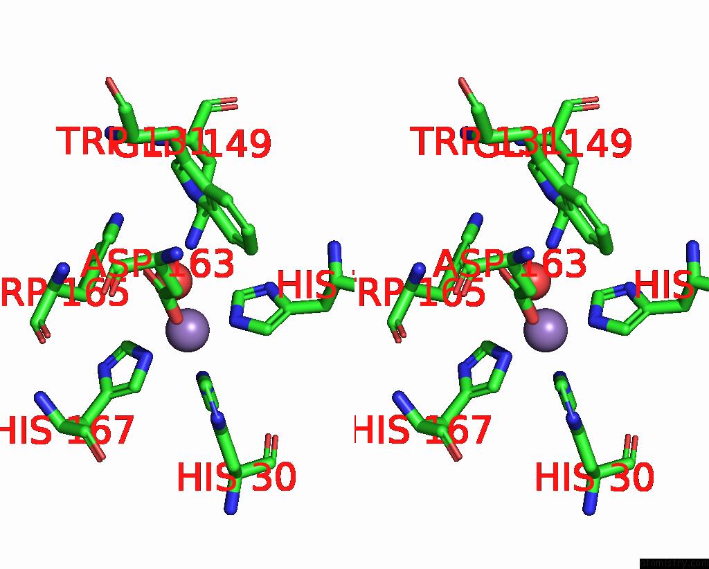
Manganese binding site 4 out of 8 in 7svs
Go back to
Manganese binding site 4 out
of 8 in the Crystal Structure Analysis of the G73A Mutant of Superoxide Dismutase From Trichoderma Reesei
Mono view
Stereo pair view
Mono view
Stereo pair view
A full contact list of Manganese with other atoms in the Mn binding
site number 4 of Crystal Structure Analysis of the G73A Mutant of Superoxide Dismutase From Trichoderma Reesei within 5.0Å range:
|
Manganese binding site 5 out of 8 in 7svs
Go back to
Manganese binding site 5 out
of 8 in the Crystal Structure Analysis of the G73A Mutant of Superoxide Dismutase From Trichoderma Reesei
Mono view
Stereo pair view
Mono view
Stereo pair view
A full contact list of Manganese with other atoms in the Mn binding
site number 5 of Crystal Structure Analysis of the G73A Mutant of Superoxide Dismutase From Trichoderma Reesei within 5.0Å range:
|
Manganese binding site 6 out of 8 in 7svs
Go back to
Manganese binding site 6 out
of 8 in the Crystal Structure Analysis of the G73A Mutant of Superoxide Dismutase From Trichoderma Reesei
Mono view

Stereo pair view
Mono view
Stereo pair view
A full contact list of Manganese with other atoms in the Mn binding
site number 6 of Crystal Structure Analysis of the G73A Mutant of Superoxide Dismutase From Trichoderma Reesei within 5.0Å range:
|
Manganese binding site 7 out of 8 in 7svs
Go back to
Manganese binding site 7 out
of 8 in the Crystal Structure Analysis of the G73A Mutant of Superoxide Dismutase From Trichoderma Reesei
Mono view

Stereo pair view
Mono view
Stereo pair view
A full contact list of Manganese with other atoms in the Mn binding
site number 7 of Crystal Structure Analysis of the G73A Mutant of Superoxide Dismutase From Trichoderma Reesei within 5.0Å range:
|
Manganese binding site 8 out of 8 in 7svs
Go back to
Manganese binding site 8 out
of 8 in the Crystal Structure Analysis of the G73A Mutant of Superoxide Dismutase From Trichoderma Reesei
Mono view
Stereo pair view
Mono view
Stereo pair view
A full contact list of Manganese with other atoms in the Mn binding
site number 8 of Crystal Structure Analysis of the G73A Mutant of Superoxide Dismutase From Trichoderma Reesei within 5.0Å range:
|
Reference:
E.Mendoza Rengifo,
L.Stelmastchuk Benassi Fontolan,
J.Ribamar Ferreira-Junior,
L.Bleicher,
J.Penner-Hahn,
R.Charles Garratt.
Unexpected Plasticity of the Quaternary Structure of Iron-Manganese Superoxide Dismutases. J.Struct.Biol. V. 214 07855 2022.
ISSN: ESSN 1095-8657
PubMed: 35390463
DOI: 10.1016/J.JSB.2022.107855
Page generated: Sun Oct 6 10:35:16 2024
ISSN: ESSN 1095-8657
PubMed: 35390463
DOI: 10.1016/J.JSB.2022.107855
Last articles
Zn in 9J0NZn in 9J0O
Zn in 9J0P
Zn in 9FJX
Zn in 9EKB
Zn in 9C0F
Zn in 9CAH
Zn in 9CH0
Zn in 9CH3
Zn in 9CH1