Manganese »
PDB 7ohl-7sck »
7qoe »
Manganese in PDB 7qoe: Structure of A Small Alarmone Hydrolase From Leptospira Levettii
Protein crystallography data
The structure of Structure of A Small Alarmone Hydrolase From Leptospira Levettii, PDB code: 7qoe
was solved by
F.Bisiak,
D.E.Brodersen,
A.Chrenkova,
with X-Ray Crystallography technique. A brief refinement statistics is given in the table below:
| Resolution Low / High (Å) | 44.14 / 1.20 |
| Space group | P 42 21 2 |
| Cell size a, b, c (Å), α, β, γ (°) | 98.708, 98.708, 40.086, 90, 90, 90 |
| R / Rfree (%) | 15.2 / 18.1 |
Manganese Binding Sites:
The binding sites of Manganese atom in the Structure of A Small Alarmone Hydrolase From Leptospira Levettii
(pdb code 7qoe). This binding sites where shown within
5.0 Angstroms radius around Manganese atom.
In total only one binding site of Manganese was determined in the Structure of A Small Alarmone Hydrolase From Leptospira Levettii, PDB code: 7qoe:
In total only one binding site of Manganese was determined in the Structure of A Small Alarmone Hydrolase From Leptospira Levettii, PDB code: 7qoe:
Manganese binding site 1 out of 1 in 7qoe
Go back to
Manganese binding site 1 out
of 1 in the Structure of A Small Alarmone Hydrolase From Leptospira Levettii

Mono view
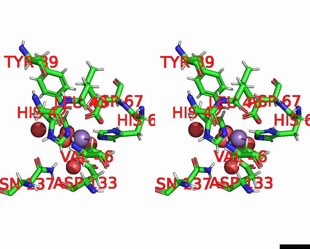
Stereo pair view
Mono view
Stereo pair view
A full contact list of Manganese with other atoms in the Mn binding
site number 1 of Structure of A Small Alarmone Hydrolase From Leptospira Levettii within 5.0Å range:
|
Reference:
F.Bisiak,
A.Chrenkova,
S.D.Zhang,
J.N.Pedersen,
D.E.Otzen,
Y.E.Zhang,
D.E.Brodersen.
Structural Variations Between Small Alarmone Hydrolase Dimers Support Different Modes of Regulation of the Stringent Response. J.Biol.Chem. V. 298 02142 2022.
ISSN: ESSN 1083-351X
PubMed: 35714769
DOI: 10.1016/J.JBC.2022.102142
Page generated: Sun Oct 6 10:27:12 2024
ISSN: ESSN 1083-351X
PubMed: 35714769
DOI: 10.1016/J.JBC.2022.102142
Last articles
Zn in 9J0NZn in 9J0O
Zn in 9J0P
Zn in 9FJX
Zn in 9EKB
Zn in 9C0F
Zn in 9CAH
Zn in 9CH0
Zn in 9CH3
Zn in 9CH1