Manganese »
PDB 7ohl-7sck »
7omb »
Manganese in PDB 7omb: Crystal Structure of Kod Dna Polymerase in A Ternary Complex with A P/T Duplex Containing An Extended 5' Single Stranded Template Overhang
Enzymatic activity of Crystal Structure of Kod Dna Polymerase in A Ternary Complex with A P/T Duplex Containing An Extended 5' Single Stranded Template Overhang
All present enzymatic activity of Crystal Structure of Kod Dna Polymerase in A Ternary Complex with A P/T Duplex Containing An Extended 5' Single Stranded Template Overhang:
2.7.7.7;
2.7.7.7;
Protein crystallography data
The structure of Crystal Structure of Kod Dna Polymerase in A Ternary Complex with A P/T Duplex Containing An Extended 5' Single Stranded Template Overhang, PDB code: 7omb
was solved by
K.Betz,
H.M.Kropp,
K.Diederichs,
A.Marx,
with X-Ray Crystallography technique. A brief refinement statistics is given in the table below:
| Resolution Low / High (Å) | 46.28 / 2.01 |
| Space group | P 21 21 2 |
| Cell size a, b, c (Å), α, β, γ (°) | 108.081, 147.153, 71.332, 90, 90, 90 |
| R / Rfree (%) | 20 / 23.6 |
Other elements in 7omb:
The structure of Crystal Structure of Kod Dna Polymerase in A Ternary Complex with A P/T Duplex Containing An Extended 5' Single Stranded Template Overhang also contains other interesting chemical elements:
| Magnesium | (Mg) | 2 atoms |
Manganese Binding Sites:
The binding sites of Manganese atom in the Crystal Structure of Kod Dna Polymerase in A Ternary Complex with A P/T Duplex Containing An Extended 5' Single Stranded Template Overhang
(pdb code 7omb). This binding sites where shown within
5.0 Angstroms radius around Manganese atom.
In total 4 binding sites of Manganese where determined in the Crystal Structure of Kod Dna Polymerase in A Ternary Complex with A P/T Duplex Containing An Extended 5' Single Stranded Template Overhang, PDB code: 7omb:
Jump to Manganese binding site number: 1; 2; 3; 4;
In total 4 binding sites of Manganese where determined in the Crystal Structure of Kod Dna Polymerase in A Ternary Complex with A P/T Duplex Containing An Extended 5' Single Stranded Template Overhang, PDB code: 7omb:
Jump to Manganese binding site number: 1; 2; 3; 4;
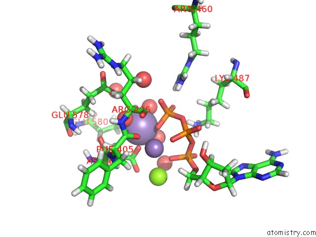
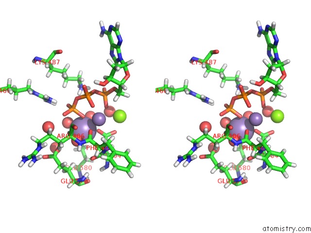
Manganese binding site 1 out of 4 in 7omb
Go back to
Manganese binding site 1 out
of 4 in the Crystal Structure of Kod Dna Polymerase in A Ternary Complex with A P/T Duplex Containing An Extended 5' Single Stranded Template Overhang
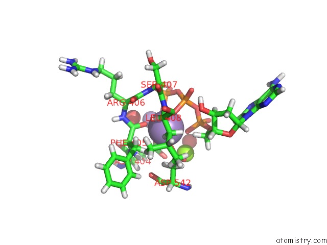
Mono view
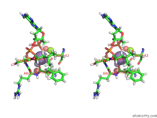
Stereo pair view
Mono view
Stereo pair view
A full contact list of Manganese with other atoms in the Mn binding
site number 1 of Crystal Structure of Kod Dna Polymerase in A Ternary Complex with A P/T Duplex Containing An Extended 5' Single Stranded Template Overhang within 5.0Å range:
|
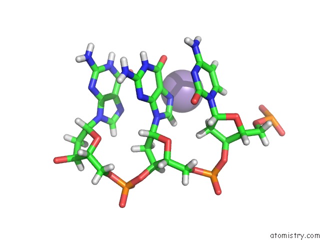
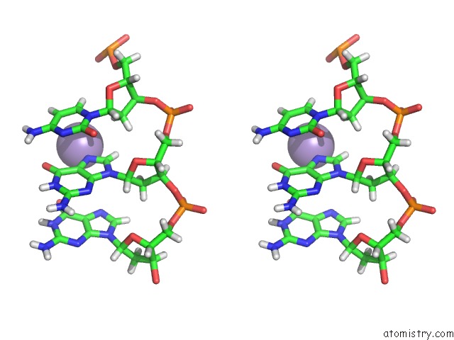
Manganese binding site 2 out of 4 in 7omb
Go back to
Manganese binding site 2 out
of 4 in the Crystal Structure of Kod Dna Polymerase in A Ternary Complex with A P/T Duplex Containing An Extended 5' Single Stranded Template Overhang
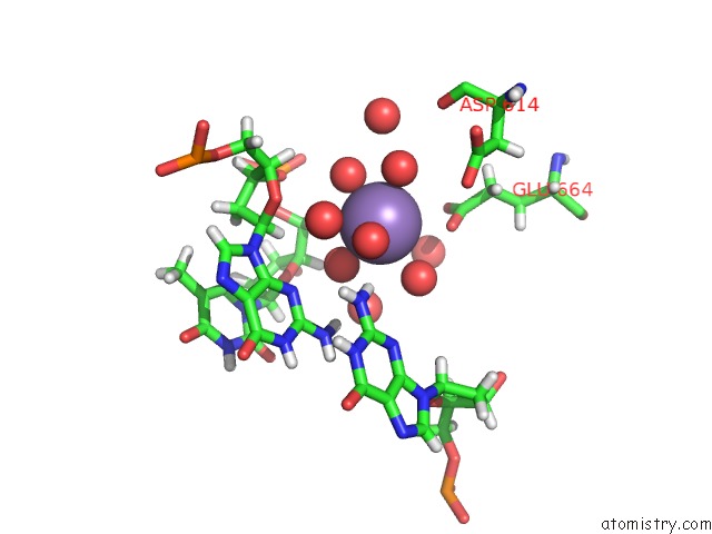
Mono view
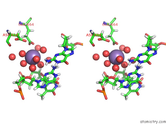
Stereo pair view
Mono view
Stereo pair view
A full contact list of Manganese with other atoms in the Mn binding
site number 2 of Crystal Structure of Kod Dna Polymerase in A Ternary Complex with A P/T Duplex Containing An Extended 5' Single Stranded Template Overhang within 5.0Å range:
|
Manganese binding site 3 out of 4 in 7omb
Go back to
Manganese binding site 3 out
of 4 in the Crystal Structure of Kod Dna Polymerase in A Ternary Complex with A P/T Duplex Containing An Extended 5' Single Stranded Template Overhang
Mono view
Stereo pair view
Mono view
Stereo pair view
A full contact list of Manganese with other atoms in the Mn binding
site number 3 of Crystal Structure of Kod Dna Polymerase in A Ternary Complex with A P/T Duplex Containing An Extended 5' Single Stranded Template Overhang within 5.0Å range:
|
Manganese binding site 4 out of 4 in 7omb
Go back to
Manganese binding site 4 out
of 4 in the Crystal Structure of Kod Dna Polymerase in A Ternary Complex with A P/T Duplex Containing An Extended 5' Single Stranded Template Overhang
Mono view
Stereo pair view
Mono view
Stereo pair view
A full contact list of Manganese with other atoms in the Mn binding
site number 4 of Crystal Structure of Kod Dna Polymerase in A Ternary Complex with A P/T Duplex Containing An Extended 5' Single Stranded Template Overhang within 5.0Å range:
|
Reference:
H.M.Kropp,
S.Ludmann,
K.Diederichs,
K.Betz,
A.Marx.
Structural Basis For the Recognition of Deaminated Nucleobases By An Archaeal Dna Polymerase. Chembiochem 2021.
ISSN: ESSN 1439-7633
PubMed: 34486208
DOI: 10.1002/CBIC.202100306
Page generated: Sun Oct 6 10:22:58 2024
ISSN: ESSN 1439-7633
PubMed: 34486208
DOI: 10.1002/CBIC.202100306
Last articles
Zn in 9J0NZn in 9J0O
Zn in 9J0P
Zn in 9FJX
Zn in 9EKB
Zn in 9C0F
Zn in 9CAH
Zn in 9CH0
Zn in 9CH3
Zn in 9CH1