Manganese »
PDB 7mxs-7ohg »
7n02 »
Manganese in PDB 7n02: X-Ray Crystallographic Structure Model of Lactococcus Lactis Prolidase Mutant D36S
Enzymatic activity of X-Ray Crystallographic Structure Model of Lactococcus Lactis Prolidase Mutant D36S
All present enzymatic activity of X-Ray Crystallographic Structure Model of Lactococcus Lactis Prolidase Mutant D36S:
3.4.13.9;
3.4.13.9;
Protein crystallography data
The structure of X-Ray Crystallographic Structure Model of Lactococcus Lactis Prolidase Mutant D36S, PDB code: 7n02
was solved by
S.Xu,
P.Grochulski,
T.Tanaka,
with X-Ray Crystallography technique. A brief refinement statistics is given in the table below:
| Resolution Low / High (Å) | 43.64 / 2.35 |
| Space group | P 21 21 21 |
| Cell size a, b, c (Å), α, β, γ (°) | 83.12, 87.27, 119.4, 90, 90, 90 |
| R / Rfree (%) | 21.2 / 27 |
Manganese Binding Sites:
The binding sites of Manganese atom in the X-Ray Crystallographic Structure Model of Lactococcus Lactis Prolidase Mutant D36S
(pdb code 7n02). This binding sites where shown within
5.0 Angstroms radius around Manganese atom.
In total 4 binding sites of Manganese where determined in the X-Ray Crystallographic Structure Model of Lactococcus Lactis Prolidase Mutant D36S, PDB code: 7n02:
Jump to Manganese binding site number: 1; 2; 3; 4;
In total 4 binding sites of Manganese where determined in the X-Ray Crystallographic Structure Model of Lactococcus Lactis Prolidase Mutant D36S, PDB code: 7n02:
Jump to Manganese binding site number: 1; 2; 3; 4;
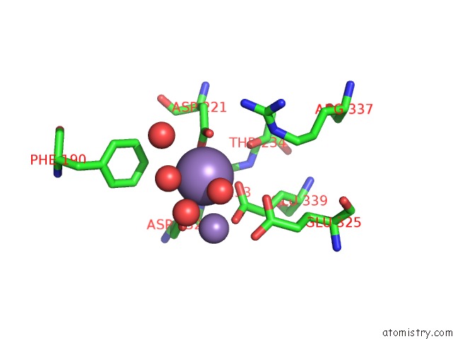
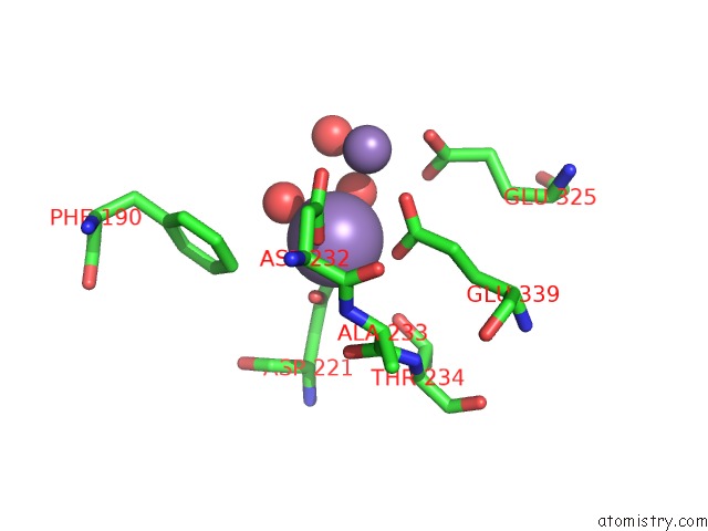
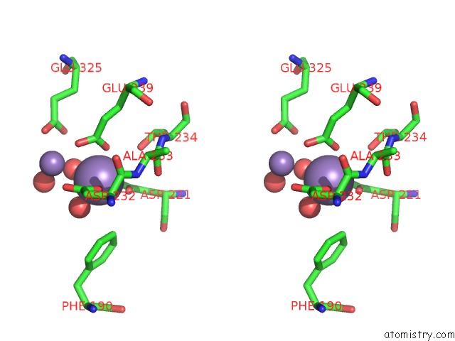
Manganese binding site 1 out of 4 in 7n02
Go back to
Manganese binding site 1 out
of 4 in the X-Ray Crystallographic Structure Model of Lactococcus Lactis Prolidase Mutant D36S
Mono view
Stereo pair view
Mono view
Stereo pair view
A full contact list of Manganese with other atoms in the Mn binding
site number 1 of X-Ray Crystallographic Structure Model of Lactococcus Lactis Prolidase Mutant D36S within 5.0Å range:
|
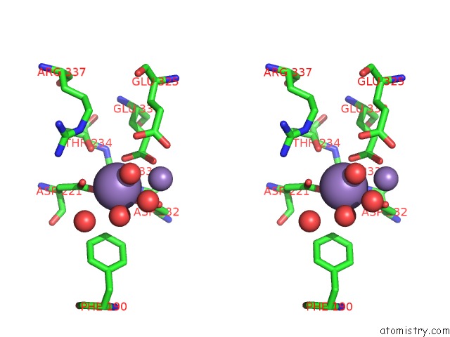
Manganese binding site 2 out of 4 in 7n02
Go back to
Manganese binding site 2 out
of 4 in the X-Ray Crystallographic Structure Model of Lactococcus Lactis Prolidase Mutant D36S
Mono view
Stereo pair view
Mono view
Stereo pair view
A full contact list of Manganese with other atoms in the Mn binding
site number 2 of X-Ray Crystallographic Structure Model of Lactococcus Lactis Prolidase Mutant D36S within 5.0Å range:
|
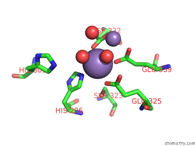
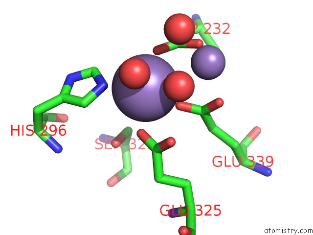
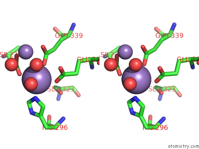
Manganese binding site 3 out of 4 in 7n02
Go back to
Manganese binding site 3 out
of 4 in the X-Ray Crystallographic Structure Model of Lactococcus Lactis Prolidase Mutant D36S
Mono view
Stereo pair view
Mono view
Stereo pair view
A full contact list of Manganese with other atoms in the Mn binding
site number 3 of X-Ray Crystallographic Structure Model of Lactococcus Lactis Prolidase Mutant D36S within 5.0Å range:
|
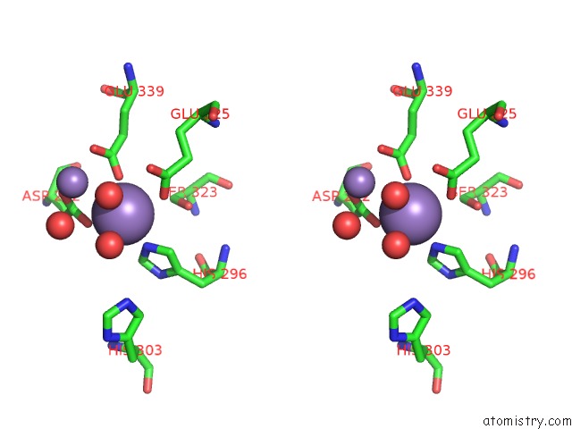
Manganese binding site 4 out of 4 in 7n02
Go back to
Manganese binding site 4 out
of 4 in the X-Ray Crystallographic Structure Model of Lactococcus Lactis Prolidase Mutant D36S
Mono view
Stereo pair view
Mono view
Stereo pair view
A full contact list of Manganese with other atoms in the Mn binding
site number 4 of X-Ray Crystallographic Structure Model of Lactococcus Lactis Prolidase Mutant D36S within 5.0Å range:
|
Reference:
O.Kgosisejo,
J.A.Chen,
P.Grochulski,
T.Tanaka.
Crystallographic Structure of Recombinant Lactococcus Lactis Prolidase to Support Proposed Structure-Function Relationships. Biochim Biophys Acta V.1865 473 2017PROTEINS Proteom.
ISSN: ISSN 1570-9639
PubMed: 28179139
DOI: 10.1016/J.BBAPAP.2017.02.004
Page generated: Sun Oct 6 10:13:10 2024
ISSN: ISSN 1570-9639
PubMed: 28179139
DOI: 10.1016/J.BBAPAP.2017.02.004
Last articles
Zn in 9JYWZn in 9IR4
Zn in 9IR3
Zn in 9GMX
Zn in 9GMW
Zn in 9JEJ
Zn in 9ERF
Zn in 9ERE
Zn in 9EGV
Zn in 9EGW