Manganese »
PDB 7lt2-7mxr »
7mvs »
Manganese in PDB 7mvs: Dna Gyrase Complexed with Uncleaved Dna and Compound 7 to 2.6A Resolution
Enzymatic activity of Dna Gyrase Complexed with Uncleaved Dna and Compound 7 to 2.6A Resolution
All present enzymatic activity of Dna Gyrase Complexed with Uncleaved Dna and Compound 7 to 2.6A Resolution:
5.6.2.2;
5.6.2.2;
Protein crystallography data
The structure of Dna Gyrase Complexed with Uncleaved Dna and Compound 7 to 2.6A Resolution, PDB code: 7mvs
was solved by
S.C.Ratigan,
C.A.Mcelroy,
with X-Ray Crystallography technique. A brief refinement statistics is given in the table below:
| Resolution Low / High (Å) | 23.74 / 2.60 |
| Space group | P 61 |
| Cell size a, b, c (Å), α, β, γ (°) | 93.017, 93.017, 406.663, 90, 90, 120 |
| R / Rfree (%) | 19.6 / 25.2 |
Other elements in 7mvs:
The structure of Dna Gyrase Complexed with Uncleaved Dna and Compound 7 to 2.6A Resolution also contains other interesting chemical elements:
| Chlorine | (Cl) | 3 atoms |
| Fluorine | (F) | 2 atoms |
Manganese Binding Sites:
The binding sites of Manganese atom in the Dna Gyrase Complexed with Uncleaved Dna and Compound 7 to 2.6A Resolution
(pdb code 7mvs). This binding sites where shown within
5.0 Angstroms radius around Manganese atom.
In total 2 binding sites of Manganese where determined in the Dna Gyrase Complexed with Uncleaved Dna and Compound 7 to 2.6A Resolution, PDB code: 7mvs:
Jump to Manganese binding site number: 1; 2;
In total 2 binding sites of Manganese where determined in the Dna Gyrase Complexed with Uncleaved Dna and Compound 7 to 2.6A Resolution, PDB code: 7mvs:
Jump to Manganese binding site number: 1; 2;
Manganese binding site 1 out of 2 in 7mvs
Go back to
Manganese binding site 1 out
of 2 in the Dna Gyrase Complexed with Uncleaved Dna and Compound 7 to 2.6A Resolution
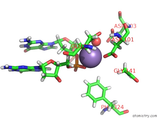
Mono view
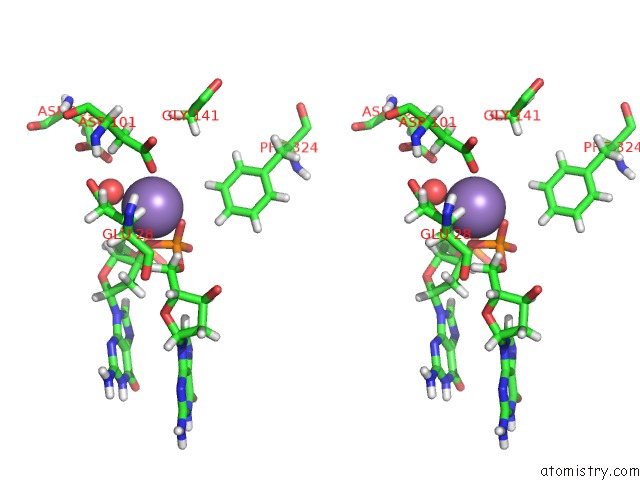
Stereo pair view
Mono view
Stereo pair view
A full contact list of Manganese with other atoms in the Mn binding
site number 1 of Dna Gyrase Complexed with Uncleaved Dna and Compound 7 to 2.6A Resolution within 5.0Å range:
|
Manganese binding site 2 out of 2 in 7mvs
Go back to
Manganese binding site 2 out
of 2 in the Dna Gyrase Complexed with Uncleaved Dna and Compound 7 to 2.6A Resolution
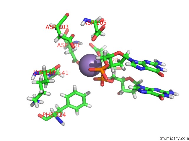
Mono view
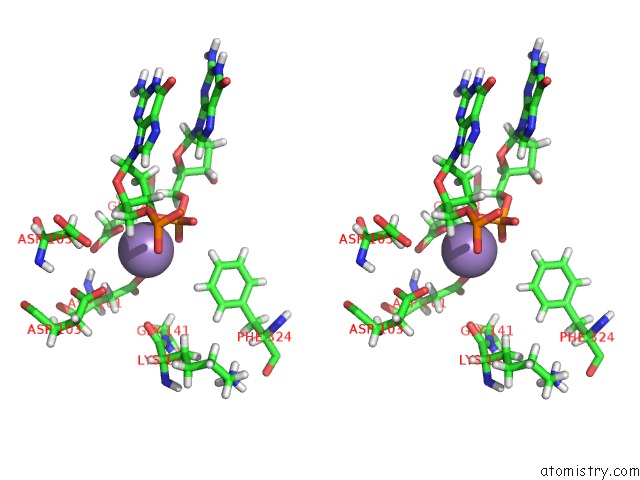
Stereo pair view
Mono view
Stereo pair view
A full contact list of Manganese with other atoms in the Mn binding
site number 2 of Dna Gyrase Complexed with Uncleaved Dna and Compound 7 to 2.6A Resolution within 5.0Å range:
|
Reference:
Y.Lu,
S.Vibhute,
L.Li,
A.Okumu,
S.C.Ratigan,
S.Nolan,
J.L.Papa,
C.A.Mann,
A.English,
A.Chen,
J.T.Seffernick,
B.Koci,
L.R.Duncan,
B.Roth,
J.E.Cummings,
R.A.Slayden,
S.Lindert,
C.A.Mcelroy,
D.J.Wozniak,
J.Yalowich,
M.J.Mitton-Fry.
Optimization of Topoiv Potency, Admet Properties, and Herg Inhibition of 5-Amino-1,3-Dioxane-Linked Novel Bacterial Topoisomerase Inhibitors: Identification of A Lead with in Vivo Efficacy Against Mrsa. J.Med.Chem. 2021.
ISSN: ISSN 0022-2623
PubMed: 34614347
DOI: 10.1021/ACS.JMEDCHEM.1C01250
Page generated: Sun Oct 6 10:09:54 2024
ISSN: ISSN 0022-2623
PubMed: 34614347
DOI: 10.1021/ACS.JMEDCHEM.1C01250
Last articles
F in 7M4PF in 7M04
F in 7M2L
F in 7M2O
F in 7M1K
F in 7M0X
F in 7M2F
F in 7M0Y
F in 7M0Z
F in 7M0M