Manganese »
PDB 7lt2-7mxr »
7mgd »
Manganese in PDB 7mgd: Concanavalin A Bound to A Dna Glycoconjugate, T(Man-T)Tt
Protein crystallography data
The structure of Concanavalin A Bound to A Dna Glycoconjugate, T(Man-T)Tt, PDB code: 7mgd
was solved by
B.E.Partridge,
P.H.Winegar,
C.A.Mirkin,
with X-Ray Crystallography technique. A brief refinement statistics is given in the table below:
| Resolution Low / High (Å) | 60.53 / 2.05 |
| Space group | P 1 21 1 |
| Cell size a, b, c (Å), α, β, γ (°) | 60.56, 63.97, 126.45, 90, 93.3, 90 |
| R / Rfree (%) | 21 / 25.5 |
Other elements in 7mgd:
The structure of Concanavalin A Bound to A Dna Glycoconjugate, T(Man-T)Tt also contains other interesting chemical elements:
| Calcium | (Ca) | 4 atoms |
Manganese Binding Sites:
The binding sites of Manganese atom in the Concanavalin A Bound to A Dna Glycoconjugate, T(Man-T)Tt
(pdb code 7mgd). This binding sites where shown within
5.0 Angstroms radius around Manganese atom.
In total 4 binding sites of Manganese where determined in the Concanavalin A Bound to A Dna Glycoconjugate, T(Man-T)Tt, PDB code: 7mgd:
Jump to Manganese binding site number: 1; 2; 3; 4;
In total 4 binding sites of Manganese where determined in the Concanavalin A Bound to A Dna Glycoconjugate, T(Man-T)Tt, PDB code: 7mgd:
Jump to Manganese binding site number: 1; 2; 3; 4;
Manganese binding site 1 out of 4 in 7mgd
Go back to
Manganese binding site 1 out
of 4 in the Concanavalin A Bound to A Dna Glycoconjugate, T(Man-T)Tt
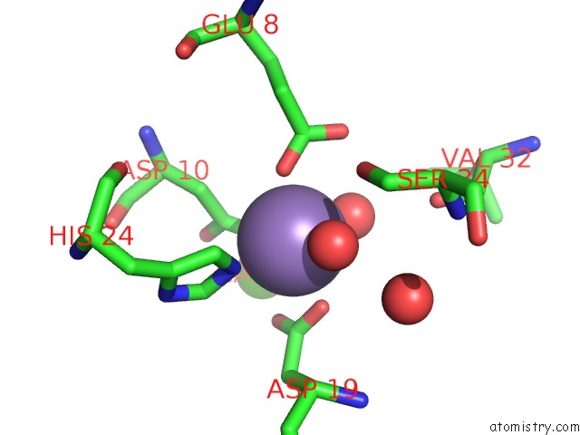
Mono view

Stereo pair view
Mono view
Stereo pair view
A full contact list of Manganese with other atoms in the Mn binding
site number 1 of Concanavalin A Bound to A Dna Glycoconjugate, T(Man-T)Tt within 5.0Å range:
|
Manganese binding site 2 out of 4 in 7mgd
Go back to
Manganese binding site 2 out
of 4 in the Concanavalin A Bound to A Dna Glycoconjugate, T(Man-T)Tt
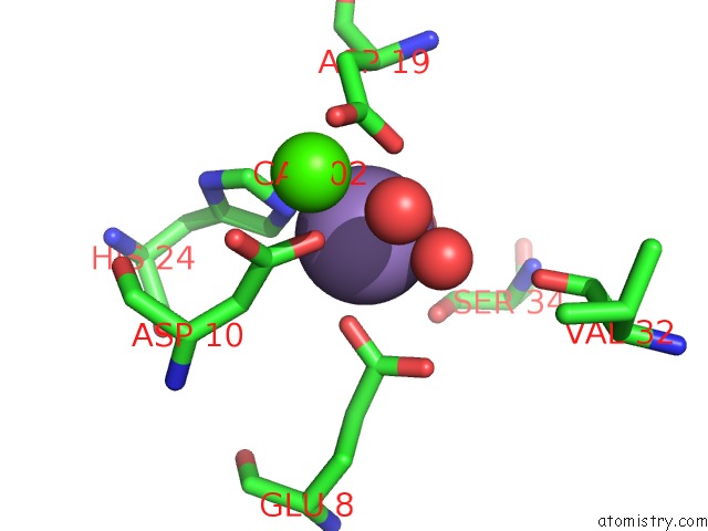
Mono view

Stereo pair view
Mono view
Stereo pair view
A full contact list of Manganese with other atoms in the Mn binding
site number 2 of Concanavalin A Bound to A Dna Glycoconjugate, T(Man-T)Tt within 5.0Å range:
|
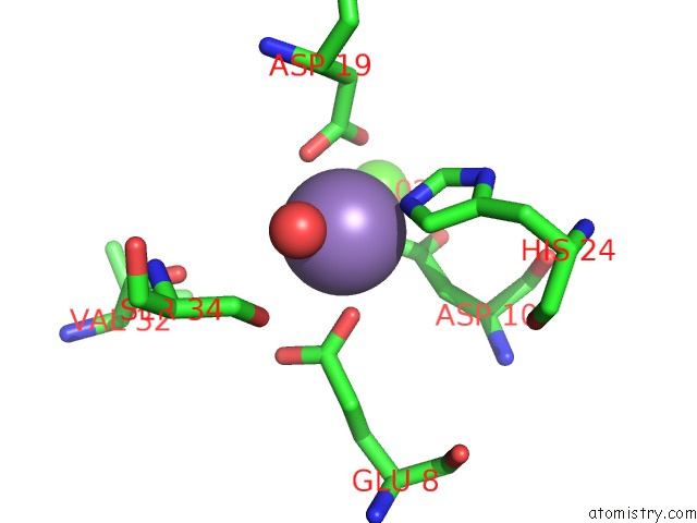
Manganese binding site 3 out of 4 in 7mgd
Go back to
Manganese binding site 3 out
of 4 in the Concanavalin A Bound to A Dna Glycoconjugate, T(Man-T)Tt

Mono view

Stereo pair view
Mono view
Stereo pair view
A full contact list of Manganese with other atoms in the Mn binding
site number 3 of Concanavalin A Bound to A Dna Glycoconjugate, T(Man-T)Tt within 5.0Å range:
|
Manganese binding site 4 out of 4 in 7mgd
Go back to
Manganese binding site 4 out
of 4 in the Concanavalin A Bound to A Dna Glycoconjugate, T(Man-T)Tt
Mono view

Stereo pair view
Mono view
Stereo pair view
A full contact list of Manganese with other atoms in the Mn binding
site number 4 of Concanavalin A Bound to A Dna Glycoconjugate, T(Man-T)Tt within 5.0Å range:
|
Reference:
B.E.Partridge,
P.H.Winegar,
Z.Han,
C.A.Mirkin.
Redefining Protein Interfaces Within Protein Single Crystals with Dna Journal of the American 2021CHEMICAL Society.
DOI: 10.1021/JACS.1C04191
Page generated: Sat Aug 16 23:18:40 2025
DOI: 10.1021/JACS.1C04191
Last articles
Na in 1VQLNa in 1VQK
Na in 1VQ9
Na in 1VQ8
Na in 1VQ7
Na in 1VQ5
Na in 1VQ6
Na in 1VQ4
Na in 1VI6
Na in 1VKG