Manganese »
PDB 7fqa-7kst »
7k3u »
Manganese in PDB 7k3u: X-Ray Crystallographic Structure Model of Lactococcus Lactis Prolidase Mutant R293S
Enzymatic activity of X-Ray Crystallographic Structure Model of Lactococcus Lactis Prolidase Mutant R293S
All present enzymatic activity of X-Ray Crystallographic Structure Model of Lactococcus Lactis Prolidase Mutant R293S:
3.4.13.9;
3.4.13.9;
Protein crystallography data
The structure of X-Ray Crystallographic Structure Model of Lactococcus Lactis Prolidase Mutant R293S, PDB code: 7k3u
was solved by
S.Xu,
P.Grochulski,
T.Tanaka,
with X-Ray Crystallography technique. A brief refinement statistics is given in the table below:
| Resolution Low / High (Å) | 49.19 / 2.70 |
| Space group | P 1 21 1 |
| Cell size a, b, c (Å), α, β, γ (°) | 146.300, 80.000, 187.150, 90.00, 102.03, 90.00 |
| R / Rfree (%) | 19.8 / 28.8 |
Manganese Binding Sites:
Pages:
>>> Page 1 <<< Page 2, Binding sites: 11 - 20;Binding sites:
The binding sites of Manganese atom in the X-Ray Crystallographic Structure Model of Lactococcus Lactis Prolidase Mutant R293S (pdb code 7k3u). This binding sites where shown within 5.0 Angstroms radius around Manganese atom.In total 20 binding sites of Manganese where determined in the X-Ray Crystallographic Structure Model of Lactococcus Lactis Prolidase Mutant R293S, PDB code: 7k3u:
Jump to Manganese binding site number: 1; 2; 3; 4; 5; 6; 7; 8; 9; 10;
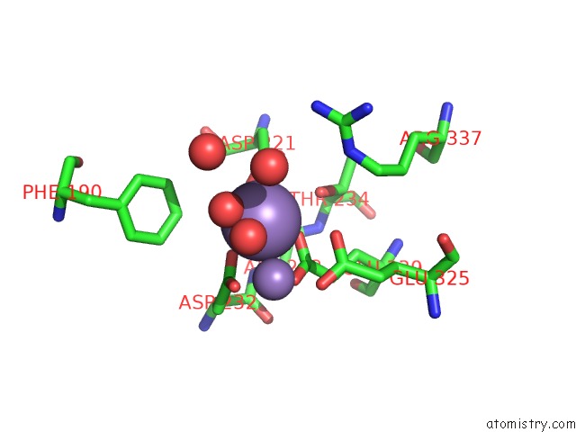
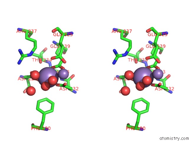
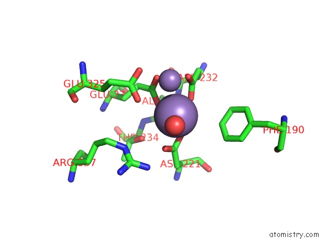
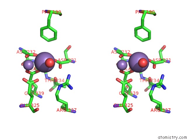
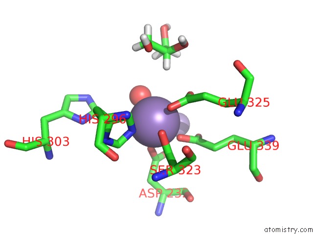
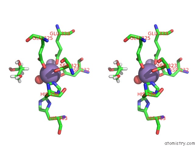
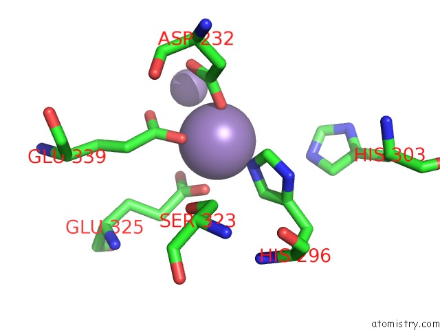
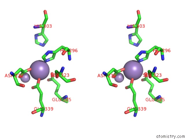
Manganese binding site 1 out of 20 in 7k3u
Go back to
Manganese binding site 1 out
of 20 in the X-Ray Crystallographic Structure Model of Lactococcus Lactis Prolidase Mutant R293S
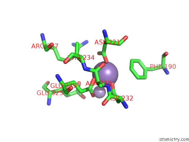
Mono view
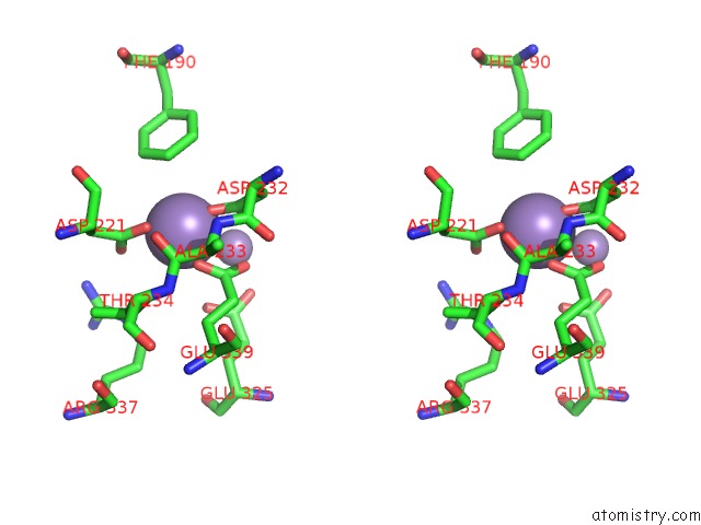
Stereo pair view
Mono view
Stereo pair view
A full contact list of Manganese with other atoms in the Mn binding
site number 1 of X-Ray Crystallographic Structure Model of Lactococcus Lactis Prolidase Mutant R293S within 5.0Å range:
|
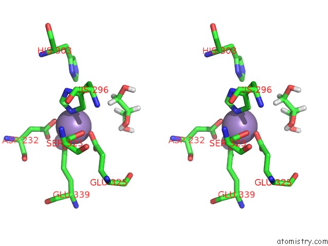
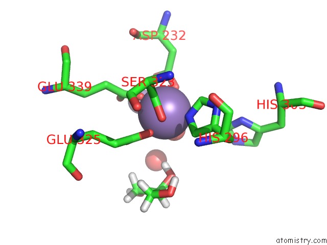
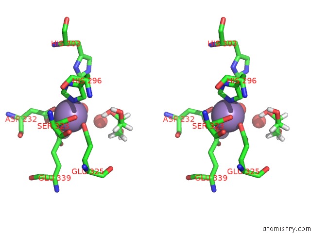
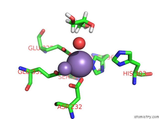
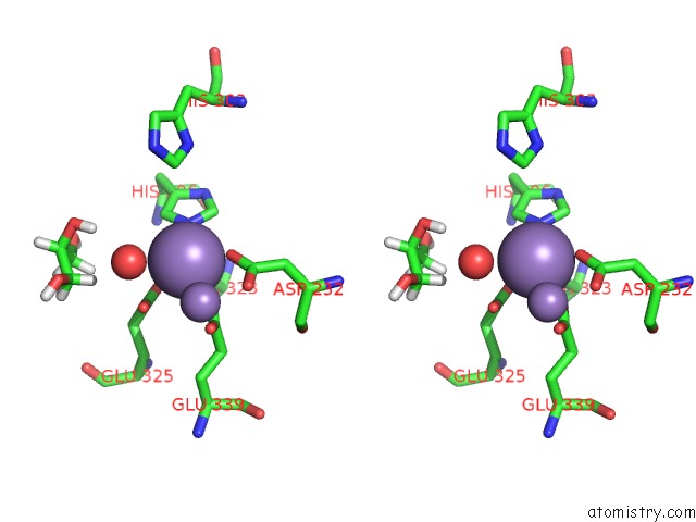
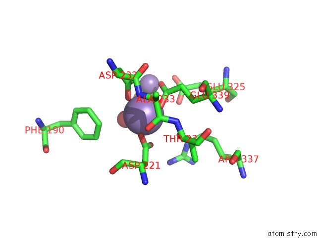
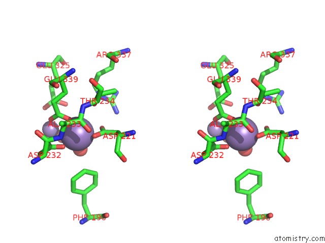
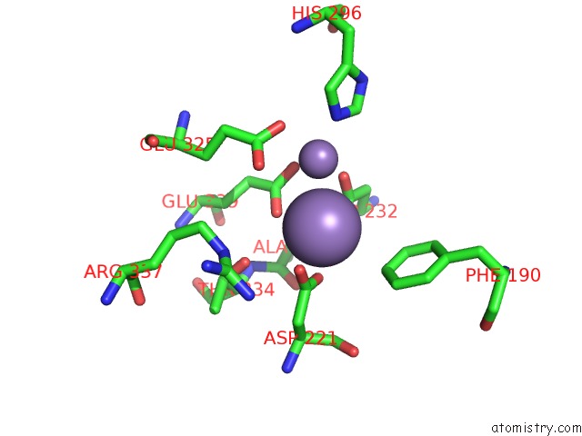
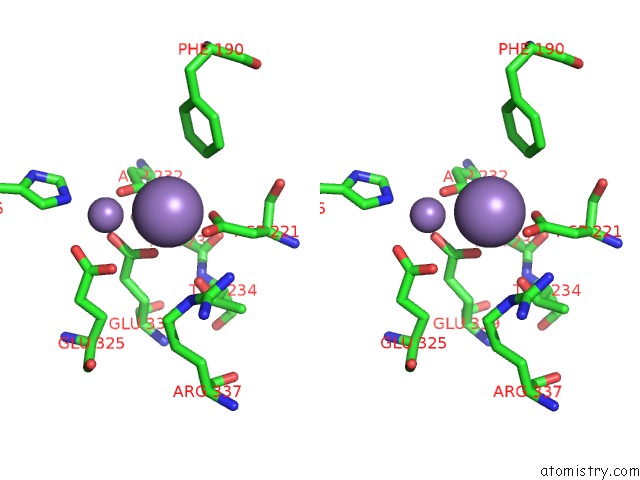
Manganese binding site 2 out of 20 in 7k3u
Go back to
Manganese binding site 2 out
of 20 in the X-Ray Crystallographic Structure Model of Lactococcus Lactis Prolidase Mutant R293S
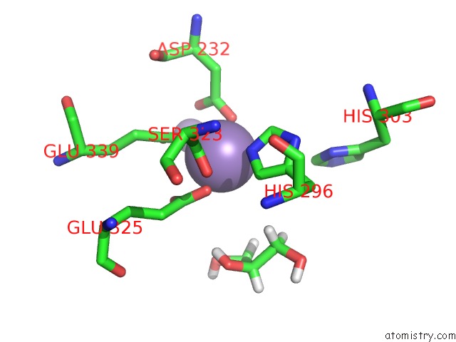
Mono view
Stereo pair view
Mono view
Stereo pair view
A full contact list of Manganese with other atoms in the Mn binding
site number 2 of X-Ray Crystallographic Structure Model of Lactococcus Lactis Prolidase Mutant R293S within 5.0Å range:
|
Manganese binding site 3 out of 20 in 7k3u
Go back to
Manganese binding site 3 out
of 20 in the X-Ray Crystallographic Structure Model of Lactococcus Lactis Prolidase Mutant R293S
Mono view
Stereo pair view
Mono view
Stereo pair view
A full contact list of Manganese with other atoms in the Mn binding
site number 3 of X-Ray Crystallographic Structure Model of Lactococcus Lactis Prolidase Mutant R293S within 5.0Å range:
|
Manganese binding site 4 out of 20 in 7k3u
Go back to
Manganese binding site 4 out
of 20 in the X-Ray Crystallographic Structure Model of Lactococcus Lactis Prolidase Mutant R293S
Mono view
Stereo pair view
Mono view
Stereo pair view
A full contact list of Manganese with other atoms in the Mn binding
site number 4 of X-Ray Crystallographic Structure Model of Lactococcus Lactis Prolidase Mutant R293S within 5.0Å range:
|
Manganese binding site 5 out of 20 in 7k3u
Go back to
Manganese binding site 5 out
of 20 in the X-Ray Crystallographic Structure Model of Lactococcus Lactis Prolidase Mutant R293S
Mono view
Stereo pair view
Mono view
Stereo pair view
A full contact list of Manganese with other atoms in the Mn binding
site number 5 of X-Ray Crystallographic Structure Model of Lactococcus Lactis Prolidase Mutant R293S within 5.0Å range:
|
Manganese binding site 6 out of 20 in 7k3u
Go back to
Manganese binding site 6 out
of 20 in the X-Ray Crystallographic Structure Model of Lactococcus Lactis Prolidase Mutant R293S
Mono view
Stereo pair view
Mono view
Stereo pair view
A full contact list of Manganese with other atoms in the Mn binding
site number 6 of X-Ray Crystallographic Structure Model of Lactococcus Lactis Prolidase Mutant R293S within 5.0Å range:
|
Manganese binding site 7 out of 20 in 7k3u
Go back to
Manganese binding site 7 out
of 20 in the X-Ray Crystallographic Structure Model of Lactococcus Lactis Prolidase Mutant R293S
Mono view
Stereo pair view
Mono view
Stereo pair view
A full contact list of Manganese with other atoms in the Mn binding
site number 7 of X-Ray Crystallographic Structure Model of Lactococcus Lactis Prolidase Mutant R293S within 5.0Å range:
|
Manganese binding site 8 out of 20 in 7k3u
Go back to
Manganese binding site 8 out
of 20 in the X-Ray Crystallographic Structure Model of Lactococcus Lactis Prolidase Mutant R293S
Mono view
Stereo pair view
Mono view
Stereo pair view
A full contact list of Manganese with other atoms in the Mn binding
site number 8 of X-Ray Crystallographic Structure Model of Lactococcus Lactis Prolidase Mutant R293S within 5.0Å range:
|
Manganese binding site 9 out of 20 in 7k3u
Go back to
Manganese binding site 9 out
of 20 in the X-Ray Crystallographic Structure Model of Lactococcus Lactis Prolidase Mutant R293S
Mono view
Stereo pair view
Mono view
Stereo pair view
A full contact list of Manganese with other atoms in the Mn binding
site number 9 of X-Ray Crystallographic Structure Model of Lactococcus Lactis Prolidase Mutant R293S within 5.0Å range:
|
Manganese binding site 10 out of 20 in 7k3u
Go back to
Manganese binding site 10 out
of 20 in the X-Ray Crystallographic Structure Model of Lactococcus Lactis Prolidase Mutant R293S
Mono view
Stereo pair view
Mono view
Stereo pair view
A full contact list of Manganese with other atoms in the Mn binding
site number 10 of X-Ray Crystallographic Structure Model of Lactococcus Lactis Prolidase Mutant R293S within 5.0Å range:
|
Reference:
O.Kgosisejo,
J.A.Chen,
P.Grochulski,
T.Tanaka.
Crystallographic Structure of Recombinant Lactococcus Lactis Prolidase to Support Proposed Structure-Function Relationships. Biochim Biophys Acta V.1865 473 2017PROTEINS Proteom.
ISSN: ISSN 1570-9639
PubMed: 28179139
DOI: 10.1016/J.BBAPAP.2017.02.004
Page generated: Sun Oct 6 09:20:51 2024
ISSN: ISSN 1570-9639
PubMed: 28179139
DOI: 10.1016/J.BBAPAP.2017.02.004
Last articles
F in 7KYTF in 7KYB
F in 7KYC
F in 7KYA
F in 7KY5
F in 7KXW
F in 7KY9
F in 7KWA
F in 7KXT
F in 7KXC