Manganese »
PDB 7ewx-7fq9 »
7ex3 »
Manganese in PDB 7ex3: Crystal Structure of Ebinur Lake Virus Cap Snatching Endonuclease in Complex with Inhibitor 3
Enzymatic activity of Crystal Structure of Ebinur Lake Virus Cap Snatching Endonuclease in Complex with Inhibitor 3
All present enzymatic activity of Crystal Structure of Ebinur Lake Virus Cap Snatching Endonuclease in Complex with Inhibitor 3:
2.7.7.48;
2.7.7.48;
Protein crystallography data
The structure of Crystal Structure of Ebinur Lake Virus Cap Snatching Endonuclease in Complex with Inhibitor 3, PDB code: 7ex3
was solved by
W.Kuang,
Z.Hu,
P.Gong,
with X-Ray Crystallography technique. A brief refinement statistics is given in the table below:
| Resolution Low / High (Å) | 35.63 / 2.25 |
| Space group | P 21 21 21 |
| Cell size a, b, c (Å), α, β, γ (°) | 40.666, 70.873, 71.253, 90, 90, 90 |
| R / Rfree (%) | 19 / 22.6 |
Other elements in 7ex3:
The structure of Crystal Structure of Ebinur Lake Virus Cap Snatching Endonuclease in Complex with Inhibitor 3 also contains other interesting chemical elements:
| Chlorine | (Cl) | 4 atoms |
| Fluorine | (F) | 1 atom |
Manganese Binding Sites:
The binding sites of Manganese atom in the Crystal Structure of Ebinur Lake Virus Cap Snatching Endonuclease in Complex with Inhibitor 3
(pdb code 7ex3). This binding sites where shown within
5.0 Angstroms radius around Manganese atom.
In total 2 binding sites of Manganese where determined in the Crystal Structure of Ebinur Lake Virus Cap Snatching Endonuclease in Complex with Inhibitor 3, PDB code: 7ex3:
Jump to Manganese binding site number: 1; 2;
In total 2 binding sites of Manganese where determined in the Crystal Structure of Ebinur Lake Virus Cap Snatching Endonuclease in Complex with Inhibitor 3, PDB code: 7ex3:
Jump to Manganese binding site number: 1; 2;
Manganese binding site 1 out of 2 in 7ex3
Go back to
Manganese binding site 1 out
of 2 in the Crystal Structure of Ebinur Lake Virus Cap Snatching Endonuclease in Complex with Inhibitor 3
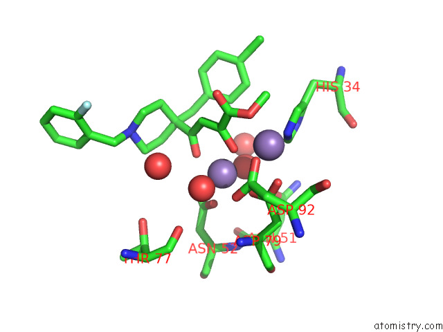
Mono view
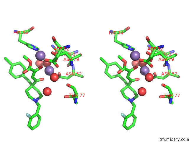
Stereo pair view
Mono view
Stereo pair view
A full contact list of Manganese with other atoms in the Mn binding
site number 1 of Crystal Structure of Ebinur Lake Virus Cap Snatching Endonuclease in Complex with Inhibitor 3 within 5.0Å range:
|
Manganese binding site 2 out of 2 in 7ex3
Go back to
Manganese binding site 2 out
of 2 in the Crystal Structure of Ebinur Lake Virus Cap Snatching Endonuclease in Complex with Inhibitor 3
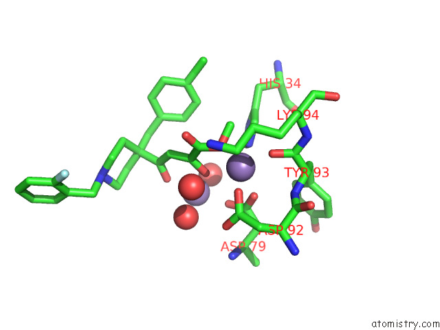
Mono view
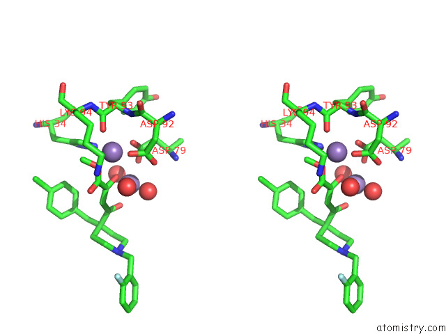
Stereo pair view
Mono view
Stereo pair view
A full contact list of Manganese with other atoms in the Mn binding
site number 2 of Crystal Structure of Ebinur Lake Virus Cap Snatching Endonuclease in Complex with Inhibitor 3 within 5.0Å range:
|
Reference:
W.Kuang,
H.Zhang,
Y.Cai,
G.Zhang,
F.Deng,
H.Li,
Y.Zhou,
M.Wang,
P.Gong,
Y.Guo,
Z.Hu.
Structural and Biochemical Basis For Development of Diketo Acid Inhibitors Targeting the Cap-Snatching Endonuclease of the Ebinur Lake Virus (Order: Bunyavirales ). J.Virol. V. 96 17321 2022.
ISSN: ESSN 1098-5514
PubMed: 35266805
DOI: 10.1128/JVI.02173-21
Page generated: Sun Oct 6 08:37:02 2024
ISSN: ESSN 1098-5514
PubMed: 35266805
DOI: 10.1128/JVI.02173-21
Last articles
Zn in 9J0NZn in 9J0O
Zn in 9J0P
Zn in 9FJX
Zn in 9EKB
Zn in 9C0F
Zn in 9CAH
Zn in 9CH0
Zn in 9CH3
Zn in 9CH1