Manganese »
PDB 7d7z-7eww »
7eul »
Manganese in PDB 7eul: Crystal Structure of C86H-H196S Mutant of N(Omega)-Hydroxy-L-Arginine Hydrolase
Enzymatic activity of Crystal Structure of C86H-H196S Mutant of N(Omega)-Hydroxy-L-Arginine Hydrolase
All present enzymatic activity of Crystal Structure of C86H-H196S Mutant of N(Omega)-Hydroxy-L-Arginine Hydrolase:
3.5.3.25;
3.5.3.25;
Protein crystallography data
The structure of Crystal Structure of C86H-H196S Mutant of N(Omega)-Hydroxy-L-Arginine Hydrolase, PDB code: 7eul
was solved by
K.Oda,
Y.Matoba,
with X-Ray Crystallography technique. A brief refinement statistics is given in the table below:
| Resolution Low / High (Å) | 41.93 / 1.45 |
| Space group | P 1 21 1 |
| Cell size a, b, c (Å), α, β, γ (°) | 42.499, 50.343, 52.941, 90, 99.42, 90 |
| R / Rfree (%) | 16.9 / 19.3 |
Other elements in 7eul:
The structure of Crystal Structure of C86H-H196S Mutant of N(Omega)-Hydroxy-L-Arginine Hydrolase also contains other interesting chemical elements:
| Magnesium | (Mg) | 2 atoms |
Manganese Binding Sites:
The binding sites of Manganese atom in the Crystal Structure of C86H-H196S Mutant of N(Omega)-Hydroxy-L-Arginine Hydrolase
(pdb code 7eul). This binding sites where shown within
5.0 Angstroms radius around Manganese atom.
In total 3 binding sites of Manganese where determined in the Crystal Structure of C86H-H196S Mutant of N(Omega)-Hydroxy-L-Arginine Hydrolase, PDB code: 7eul:
Jump to Manganese binding site number: 1; 2; 3;
In total 3 binding sites of Manganese where determined in the Crystal Structure of C86H-H196S Mutant of N(Omega)-Hydroxy-L-Arginine Hydrolase, PDB code: 7eul:
Jump to Manganese binding site number: 1; 2; 3;
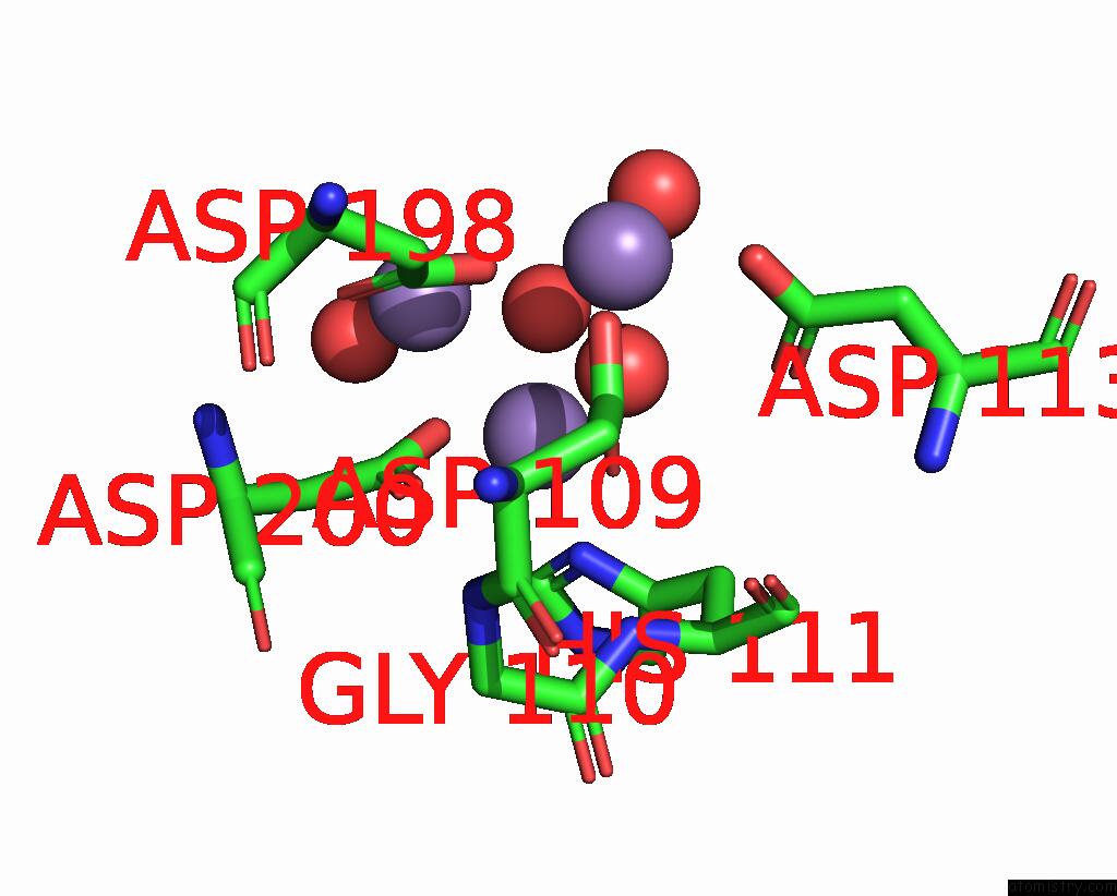
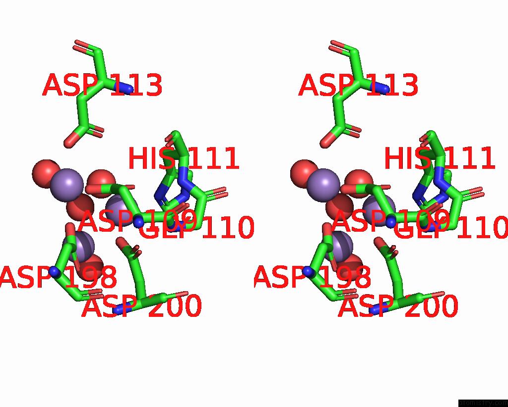
Manganese binding site 1 out of 3 in 7eul
Go back to
Manganese binding site 1 out
of 3 in the Crystal Structure of C86H-H196S Mutant of N(Omega)-Hydroxy-L-Arginine Hydrolase
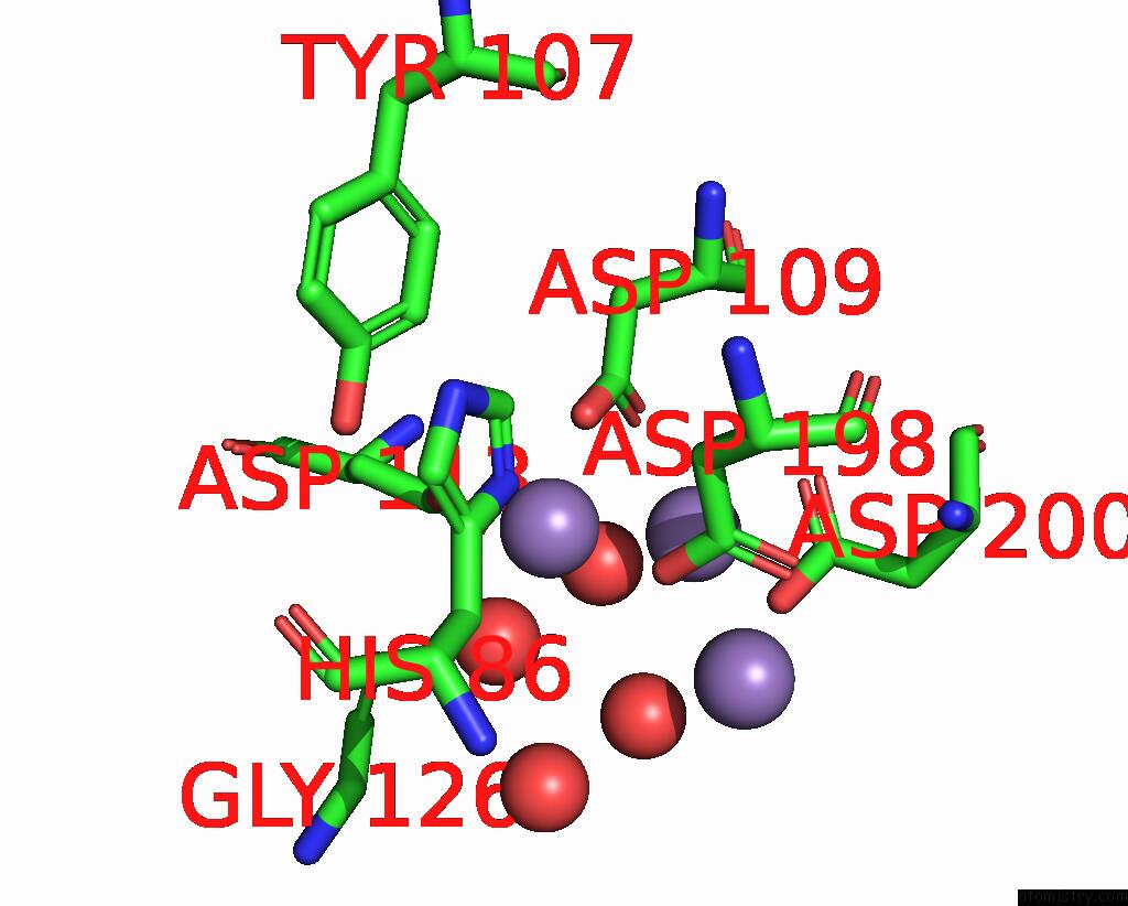
Mono view
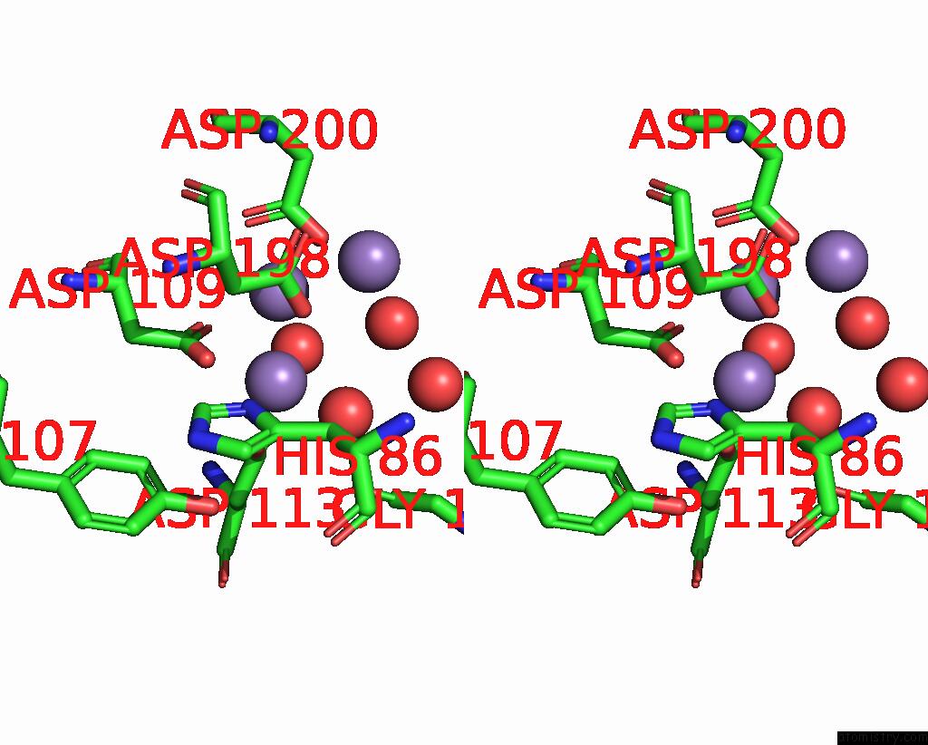
Stereo pair view
Mono view
Stereo pair view
A full contact list of Manganese with other atoms in the Mn binding
site number 1 of Crystal Structure of C86H-H196S Mutant of N(Omega)-Hydroxy-L-Arginine Hydrolase within 5.0Å range:
|
Manganese binding site 2 out of 3 in 7eul
Go back to
Manganese binding site 2 out
of 3 in the Crystal Structure of C86H-H196S Mutant of N(Omega)-Hydroxy-L-Arginine Hydrolase
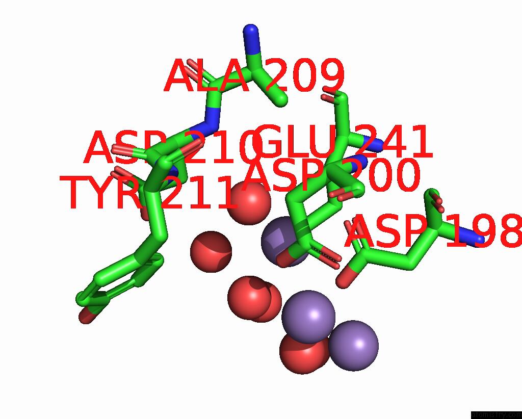
Mono view
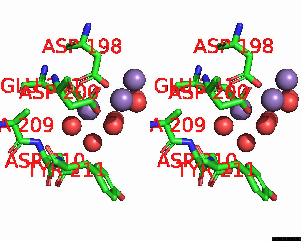
Stereo pair view
Mono view
Stereo pair view
A full contact list of Manganese with other atoms in the Mn binding
site number 2 of Crystal Structure of C86H-H196S Mutant of N(Omega)-Hydroxy-L-Arginine Hydrolase within 5.0Å range:
|
Manganese binding site 3 out of 3 in 7eul
Go back to
Manganese binding site 3 out
of 3 in the Crystal Structure of C86H-H196S Mutant of N(Omega)-Hydroxy-L-Arginine Hydrolase
Mono view
Stereo pair view
Mono view
Stereo pair view
A full contact list of Manganese with other atoms in the Mn binding
site number 3 of Crystal Structure of C86H-H196S Mutant of N(Omega)-Hydroxy-L-Arginine Hydrolase within 5.0Å range:
|
Reference:
K.Oda,
T.Sakaguchi,
Y.Matoba.
Catalytic Mechanism of Dcsb: Arginase Framework Used For Hydrolyzing Its Inhibitor. Protein Sci. V. 31 E4338 2022.
ISSN: ESSN 1469-896X
PubMed: 35634777
DOI: 10.1002/PRO.4338
Page generated: Sun Oct 6 08:34:18 2024
ISSN: ESSN 1469-896X
PubMed: 35634777
DOI: 10.1002/PRO.4338
Last articles
Zn in 9J0NZn in 9J0O
Zn in 9J0P
Zn in 9FJX
Zn in 9EKB
Zn in 9C0F
Zn in 9CAH
Zn in 9CH0
Zn in 9CH3
Zn in 9CH1