Manganese »
PDB 7d7z-7eww »
7e9s »
Manganese in PDB 7e9s: Archaeal Oligosaccharyltransferase Aglb From Archaeoglobus Fulgidus in Complex with An Inhibitory Peptide and A Dolichol-Phosphate
Enzymatic activity of Archaeal Oligosaccharyltransferase Aglb From Archaeoglobus Fulgidus in Complex with An Inhibitory Peptide and A Dolichol-Phosphate
All present enzymatic activity of Archaeal Oligosaccharyltransferase Aglb From Archaeoglobus Fulgidus in Complex with An Inhibitory Peptide and A Dolichol-Phosphate:
2.4.99.21;
2.4.99.21;
Protein crystallography data
The structure of Archaeal Oligosaccharyltransferase Aglb From Archaeoglobus Fulgidus in Complex with An Inhibitory Peptide and A Dolichol-Phosphate, PDB code: 7e9s
was solved by
Y.Taguchi,
K.Hirata,
D.Kohda,
with X-Ray Crystallography technique. A brief refinement statistics is given in the table below:
| Resolution Low / High (Å) | 24.84 / 2.70 |
| Space group | P 21 21 2 |
| Cell size a, b, c (Å), α, β, γ (°) | 345.74, 48.69, 63.56, 90, 90, 90 |
| R / Rfree (%) | 21.5 / 25.2 |
Manganese Binding Sites:
The binding sites of Manganese atom in the Archaeal Oligosaccharyltransferase Aglb From Archaeoglobus Fulgidus in Complex with An Inhibitory Peptide and A Dolichol-Phosphate
(pdb code 7e9s). This binding sites where shown within
5.0 Angstroms radius around Manganese atom.
In total only one binding site of Manganese was determined in the Archaeal Oligosaccharyltransferase Aglb From Archaeoglobus Fulgidus in Complex with An Inhibitory Peptide and A Dolichol-Phosphate, PDB code: 7e9s:
In total only one binding site of Manganese was determined in the Archaeal Oligosaccharyltransferase Aglb From Archaeoglobus Fulgidus in Complex with An Inhibitory Peptide and A Dolichol-Phosphate, PDB code: 7e9s:
Manganese binding site 1 out of 1 in 7e9s
Go back to
Manganese binding site 1 out
of 1 in the Archaeal Oligosaccharyltransferase Aglb From Archaeoglobus Fulgidus in Complex with An Inhibitory Peptide and A Dolichol-Phosphate

Mono view
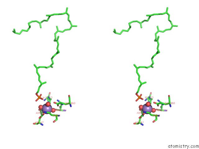
Stereo pair view
Mono view
Stereo pair view
A full contact list of Manganese with other atoms in the Mn binding
site number 1 of Archaeal Oligosaccharyltransferase Aglb From Archaeoglobus Fulgidus in Complex with An Inhibitory Peptide and A Dolichol-Phosphate within 5.0Å range:
|
Reference:
Y.Taguchi,
T.Yamasaki,
M.Ishikawa,
Y.Kawasaki,
R.Yukimura,
M.Mitani,
K.Hirata,
D.Kohda.
The Structure of An Archaeal Oligosaccharyltransferase Provides Insight Into the Strict Exclusion of Proline From the N-Glycosylation Sequon. Commun Biol V. 4 941 2021.
ISSN: ESSN 2399-3642
PubMed: 34354228
DOI: 10.1038/S42003-021-02473-8
Page generated: Sun Oct 6 08:29:17 2024
ISSN: ESSN 2399-3642
PubMed: 34354228
DOI: 10.1038/S42003-021-02473-8
Last articles
Zn in 9J0NZn in 9J0O
Zn in 9J0P
Zn in 9FJX
Zn in 9EKB
Zn in 9C0F
Zn in 9CAH
Zn in 9CH0
Zn in 9CH3
Zn in 9CH1