Manganese »
PDB 7d7z-7eww »
7d82 »
Manganese in PDB 7d82: Crystal Structure of the DOMAIN2 of Nad+ Riboswitch with Nicotinamide Adenine Dinucleotide (Nad+), Soaked in MN2+
Protein crystallography data
The structure of Crystal Structure of the DOMAIN2 of Nad+ Riboswitch with Nicotinamide Adenine Dinucleotide (Nad+), Soaked in MN2+, PDB code: 7d82
was solved by
H.Chen,
A.M.Ren,
with X-Ray Crystallography technique. A brief refinement statistics is given in the table below:
| Resolution Low / High (Å) | 27.52 / 2.49 |
| Space group | P 32 2 1 |
| Cell size a, b, c (Å), α, β, γ (°) | 75.916, 75.916, 50.289, 90.00, 90.00, 120.00 |
| R / Rfree (%) | 22.1 / 27.1 |
Other elements in 7d82:
The structure of Crystal Structure of the DOMAIN2 of Nad+ Riboswitch with Nicotinamide Adenine Dinucleotide (Nad+), Soaked in MN2+ also contains other interesting chemical elements:
| Magnesium | (Mg) | 7 atoms |
Manganese Binding Sites:
The binding sites of Manganese atom in the Crystal Structure of the DOMAIN2 of Nad+ Riboswitch with Nicotinamide Adenine Dinucleotide (Nad+), Soaked in MN2+
(pdb code 7d82). This binding sites where shown within
5.0 Angstroms radius around Manganese atom.
In total 7 binding sites of Manganese where determined in the Crystal Structure of the DOMAIN2 of Nad+ Riboswitch with Nicotinamide Adenine Dinucleotide (Nad+), Soaked in MN2+, PDB code: 7d82:
Jump to Manganese binding site number: 1; 2; 3; 4; 5; 6; 7;
In total 7 binding sites of Manganese where determined in the Crystal Structure of the DOMAIN2 of Nad+ Riboswitch with Nicotinamide Adenine Dinucleotide (Nad+), Soaked in MN2+, PDB code: 7d82:
Jump to Manganese binding site number: 1; 2; 3; 4; 5; 6; 7;
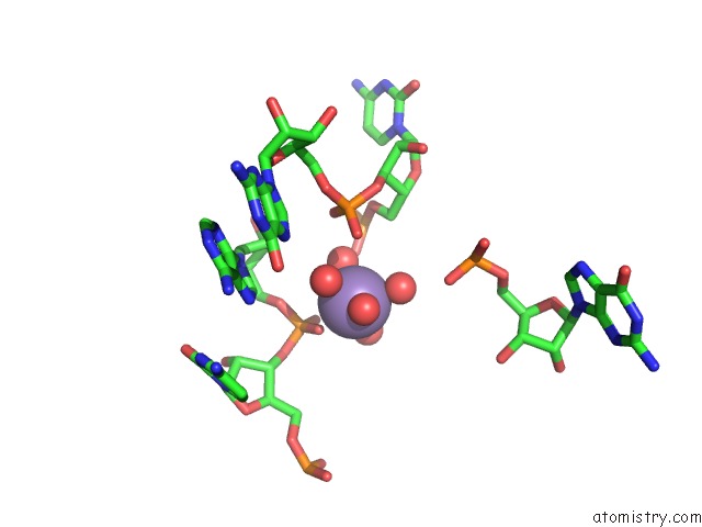
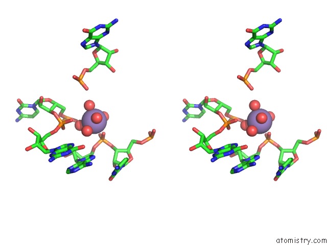
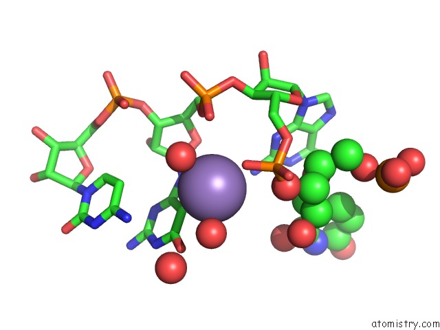
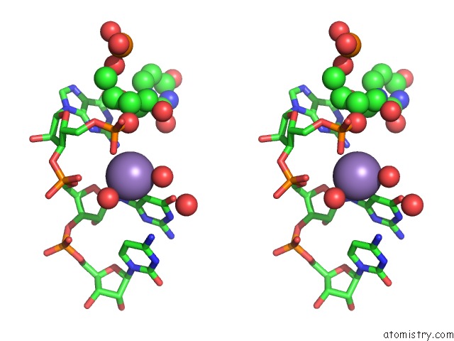
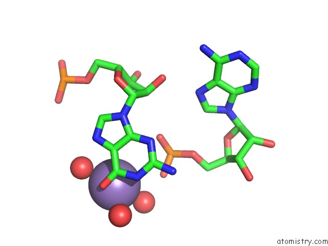
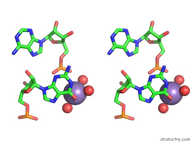
Manganese binding site 1 out of 7 in 7d82
Go back to
Manganese binding site 1 out
of 7 in the Crystal Structure of the DOMAIN2 of Nad+ Riboswitch with Nicotinamide Adenine Dinucleotide (Nad+), Soaked in MN2+
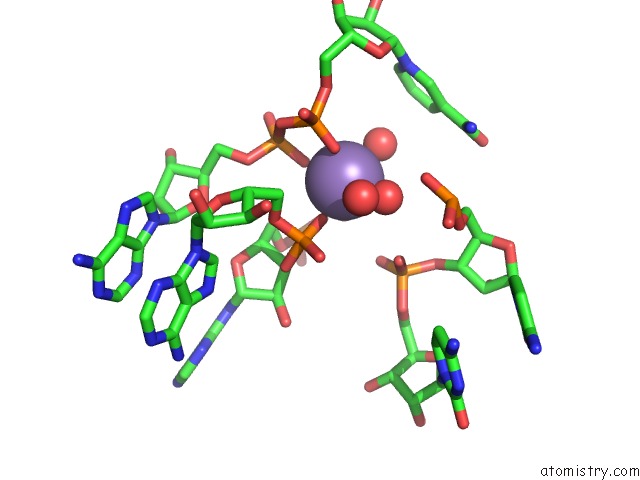
Mono view
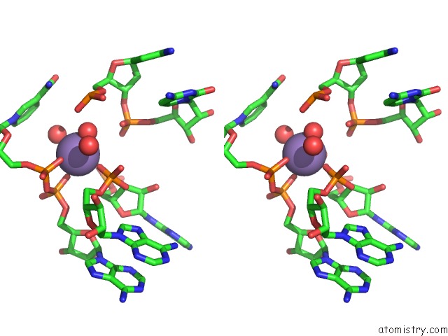
Stereo pair view
Mono view
Stereo pair view
A full contact list of Manganese with other atoms in the Mn binding
site number 1 of Crystal Structure of the DOMAIN2 of Nad+ Riboswitch with Nicotinamide Adenine Dinucleotide (Nad+), Soaked in MN2+ within 5.0Å range:
|
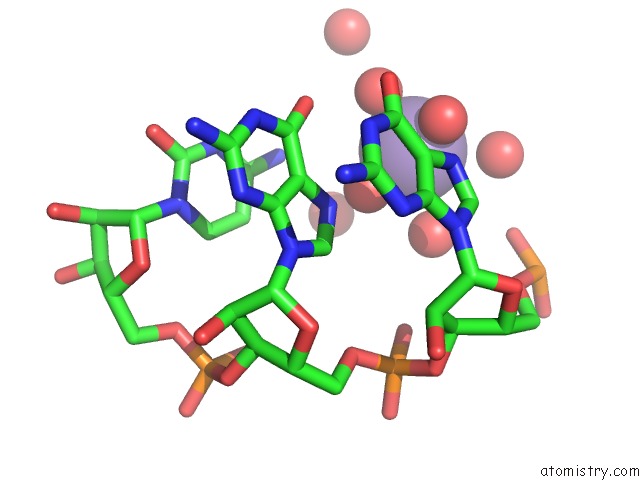
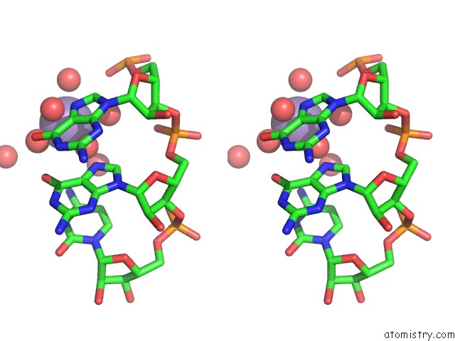
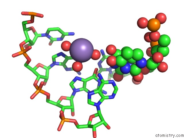
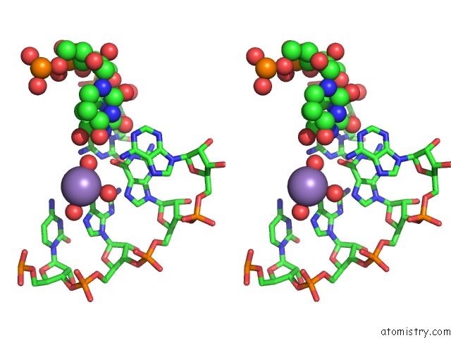
Manganese binding site 2 out of 7 in 7d82
Go back to
Manganese binding site 2 out
of 7 in the Crystal Structure of the DOMAIN2 of Nad+ Riboswitch with Nicotinamide Adenine Dinucleotide (Nad+), Soaked in MN2+
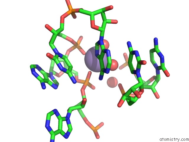
Mono view
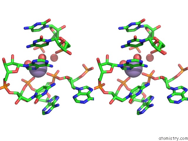
Stereo pair view
Mono view
Stereo pair view
A full contact list of Manganese with other atoms in the Mn binding
site number 2 of Crystal Structure of the DOMAIN2 of Nad+ Riboswitch with Nicotinamide Adenine Dinucleotide (Nad+), Soaked in MN2+ within 5.0Å range:
|
Manganese binding site 3 out of 7 in 7d82
Go back to
Manganese binding site 3 out
of 7 in the Crystal Structure of the DOMAIN2 of Nad+ Riboswitch with Nicotinamide Adenine Dinucleotide (Nad+), Soaked in MN2+
Mono view
Stereo pair view
Mono view
Stereo pair view
A full contact list of Manganese with other atoms in the Mn binding
site number 3 of Crystal Structure of the DOMAIN2 of Nad+ Riboswitch with Nicotinamide Adenine Dinucleotide (Nad+), Soaked in MN2+ within 5.0Å range:
|
Manganese binding site 4 out of 7 in 7d82
Go back to
Manganese binding site 4 out
of 7 in the Crystal Structure of the DOMAIN2 of Nad+ Riboswitch with Nicotinamide Adenine Dinucleotide (Nad+), Soaked in MN2+
Mono view
Stereo pair view
Mono view
Stereo pair view
A full contact list of Manganese with other atoms in the Mn binding
site number 4 of Crystal Structure of the DOMAIN2 of Nad+ Riboswitch with Nicotinamide Adenine Dinucleotide (Nad+), Soaked in MN2+ within 5.0Å range:
|
Manganese binding site 5 out of 7 in 7d82
Go back to
Manganese binding site 5 out
of 7 in the Crystal Structure of the DOMAIN2 of Nad+ Riboswitch with Nicotinamide Adenine Dinucleotide (Nad+), Soaked in MN2+
Mono view
Stereo pair view
Mono view
Stereo pair view
A full contact list of Manganese with other atoms in the Mn binding
site number 5 of Crystal Structure of the DOMAIN2 of Nad+ Riboswitch with Nicotinamide Adenine Dinucleotide (Nad+), Soaked in MN2+ within 5.0Å range:
|
Manganese binding site 6 out of 7 in 7d82
Go back to
Manganese binding site 6 out
of 7 in the Crystal Structure of the DOMAIN2 of Nad+ Riboswitch with Nicotinamide Adenine Dinucleotide (Nad+), Soaked in MN2+
Mono view
Stereo pair view
Mono view
Stereo pair view
A full contact list of Manganese with other atoms in the Mn binding
site number 6 of Crystal Structure of the DOMAIN2 of Nad+ Riboswitch with Nicotinamide Adenine Dinucleotide (Nad+), Soaked in MN2+ within 5.0Å range:
|
Manganese binding site 7 out of 7 in 7d82
Go back to
Manganese binding site 7 out
of 7 in the Crystal Structure of the DOMAIN2 of Nad+ Riboswitch with Nicotinamide Adenine Dinucleotide (Nad+), Soaked in MN2+
Mono view
Stereo pair view
Mono view
Stereo pair view
A full contact list of Manganese with other atoms in the Mn binding
site number 7 of Crystal Structure of the DOMAIN2 of Nad+ Riboswitch with Nicotinamide Adenine Dinucleotide (Nad+), Soaked in MN2+ within 5.0Å range:
|
Reference:
H.Chen,
M.Egger,
X.Xu,
L.Flemmich,
O.Krasheninina,
A.Sun,
R.Micura,
A.Ren.
Structural Distinctions Between Nad+ Riboswitch Domains 1 and 2 Determine Differential Folding and Ligand Binding. Nucleic Acids Res. 2020.
ISSN: ESSN 1362-4962
PubMed: 33170270
DOI: 10.1093/NAR/GKAA1029
Page generated: Sun Oct 6 08:26:01 2024
ISSN: ESSN 1362-4962
PubMed: 33170270
DOI: 10.1093/NAR/GKAA1029
Last articles
Fe in 2YXOFe in 2YRS
Fe in 2YXC
Fe in 2YNM
Fe in 2YVJ
Fe in 2YP1
Fe in 2YU2
Fe in 2YU1
Fe in 2YQB
Fe in 2YOO