Manganese »
PDB 6zbn-7bm0 »
7a13 »
Manganese in PDB 7a13: Crystal Structure of Human Methionine Aminopeptidase-2 in Complex with An Inhibitor GSK1978537A (Compound 27)
Enzymatic activity of Crystal Structure of Human Methionine Aminopeptidase-2 in Complex with An Inhibitor GSK1978537A (Compound 27)
All present enzymatic activity of Crystal Structure of Human Methionine Aminopeptidase-2 in Complex with An Inhibitor GSK1978537A (Compound 27):
3.4.11.18;
3.4.11.18;
Protein crystallography data
The structure of Crystal Structure of Human Methionine Aminopeptidase-2 in Complex with An Inhibitor GSK1978537A (Compound 27), PDB code: 7a13
was solved by
J.H.Thorpe,
with X-Ray Crystallography technique. A brief refinement statistics is given in the table below:
| Resolution Low / High (Å) | 29.97 / 2.04 |
| Space group | C 2 2 21 |
| Cell size a, b, c (Å), α, β, γ (°) | 89.195, 100.770, 99.396, 90.00, 90.00, 90.00 |
| R / Rfree (%) | 16.6 / 19.7 |
Other elements in 7a13:
The structure of Crystal Structure of Human Methionine Aminopeptidase-2 in Complex with An Inhibitor GSK1978537A (Compound 27) also contains other interesting chemical elements:
| Chlorine | (Cl) | 1 atom |
Manganese Binding Sites:
The binding sites of Manganese atom in the Crystal Structure of Human Methionine Aminopeptidase-2 in Complex with An Inhibitor GSK1978537A (Compound 27)
(pdb code 7a13). This binding sites where shown within
5.0 Angstroms radius around Manganese atom.
In total 2 binding sites of Manganese where determined in the Crystal Structure of Human Methionine Aminopeptidase-2 in Complex with An Inhibitor GSK1978537A (Compound 27), PDB code: 7a13:
Jump to Manganese binding site number: 1; 2;
In total 2 binding sites of Manganese where determined in the Crystal Structure of Human Methionine Aminopeptidase-2 in Complex with An Inhibitor GSK1978537A (Compound 27), PDB code: 7a13:
Jump to Manganese binding site number: 1; 2;
Manganese binding site 1 out of 2 in 7a13
Go back to
Manganese binding site 1 out
of 2 in the Crystal Structure of Human Methionine Aminopeptidase-2 in Complex with An Inhibitor GSK1978537A (Compound 27)
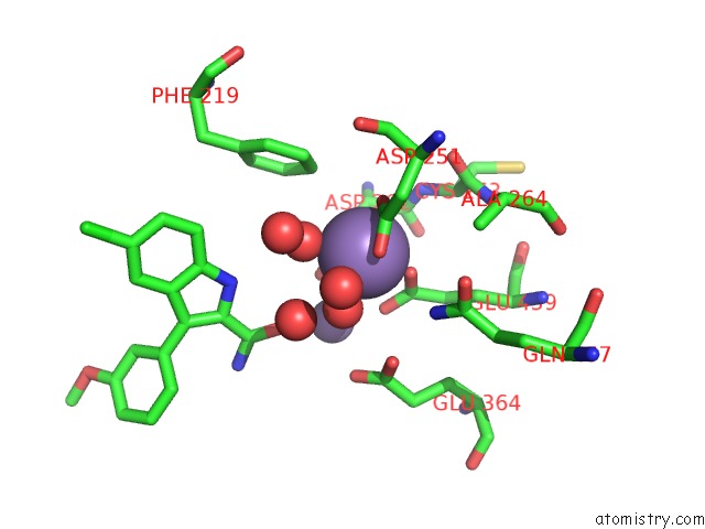
Mono view
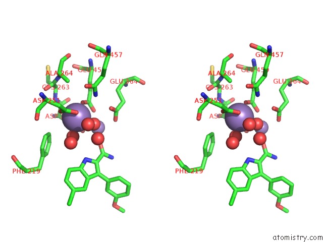
Stereo pair view
Mono view
Stereo pair view
A full contact list of Manganese with other atoms in the Mn binding
site number 1 of Crystal Structure of Human Methionine Aminopeptidase-2 in Complex with An Inhibitor GSK1978537A (Compound 27) within 5.0Å range:
|
Manganese binding site 2 out of 2 in 7a13
Go back to
Manganese binding site 2 out
of 2 in the Crystal Structure of Human Methionine Aminopeptidase-2 in Complex with An Inhibitor GSK1978537A (Compound 27)
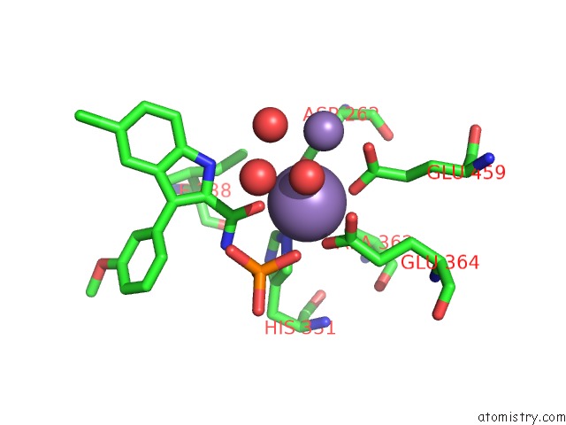
Mono view
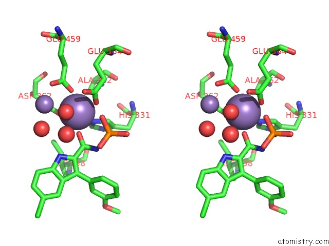
Stereo pair view
Mono view
Stereo pair view
A full contact list of Manganese with other atoms in the Mn binding
site number 2 of Crystal Structure of Human Methionine Aminopeptidase-2 in Complex with An Inhibitor GSK1978537A (Compound 27) within 5.0Å range:
|
Reference:
D.J.Hirst,
M.Brandt,
G.Bruton,
E.Christodoulou,
L.Cutler,
N.Deeks,
J.D.Goodacre,
T.Jack,
M.Lindon,
A.Miah,
K.Page,
N.Parr,
L.Shukla,
M.Sims,
P.Thomas,
J.Thorpe,
D.S.Holmes.
Structure-Based Optimisation of Orally Active & Reversible Metap-2 Inhibitors Maintaining A Tight 'Molecular Budget'. Bioorg.Med.Chem.Lett. V. 30 27533 2020.
ISSN: ESSN 1464-3405
PubMed: 32919012
DOI: 10.1016/J.BMCL.2020.127533
Page generated: Sun Oct 6 08:01:15 2024
ISSN: ESSN 1464-3405
PubMed: 32919012
DOI: 10.1016/J.BMCL.2020.127533
Last articles
Zn in 9J0NZn in 9J0O
Zn in 9J0P
Zn in 9FJX
Zn in 9EKB
Zn in 9C0F
Zn in 9CAH
Zn in 9CH0
Zn in 9CH3
Zn in 9CH1