Manganese »
PDB 6wj4-6z6r »
6y2n »
Manganese in PDB 6y2n: Crystal Structure of Ribonucleotide Reductase R2 Subunit Solved By Serial Synchrotron Crystallography
Enzymatic activity of Crystal Structure of Ribonucleotide Reductase R2 Subunit Solved By Serial Synchrotron Crystallography
All present enzymatic activity of Crystal Structure of Ribonucleotide Reductase R2 Subunit Solved By Serial Synchrotron Crystallography:
1.17.4.1;
1.17.4.1;
Protein crystallography data
The structure of Crystal Structure of Ribonucleotide Reductase R2 Subunit Solved By Serial Synchrotron Crystallography, PDB code: 6y2n
was solved by
A.Shilova,
H.Lebrette,
O.Aurelius,
M.Hogbom,
U.Mueller,
with X-Ray Crystallography technique. A brief refinement statistics is given in the table below:
| Resolution Low / High (Å) | 59.30 / 2.40 |
| Space group | P 41 21 2 |
| Cell size a, b, c (Å), α, β, γ (°) | 64.320, 64.320, 153.150, 90.00, 90.00, 90.00 |
| R / Rfree (%) | 17.3 / 22.1 |
Other elements in 6y2n:
The structure of Crystal Structure of Ribonucleotide Reductase R2 Subunit Solved By Serial Synchrotron Crystallography also contains other interesting chemical elements:
| Iron | (Fe) | 1 atom |
Manganese Binding Sites:
The binding sites of Manganese atom in the Crystal Structure of Ribonucleotide Reductase R2 Subunit Solved By Serial Synchrotron Crystallography
(pdb code 6y2n). This binding sites where shown within
5.0 Angstroms radius around Manganese atom.
In total only one binding site of Manganese was determined in the Crystal Structure of Ribonucleotide Reductase R2 Subunit Solved By Serial Synchrotron Crystallography, PDB code: 6y2n:
In total only one binding site of Manganese was determined in the Crystal Structure of Ribonucleotide Reductase R2 Subunit Solved By Serial Synchrotron Crystallography, PDB code: 6y2n:
Manganese binding site 1 out of 1 in 6y2n
Go back to
Manganese binding site 1 out
of 1 in the Crystal Structure of Ribonucleotide Reductase R2 Subunit Solved By Serial Synchrotron Crystallography
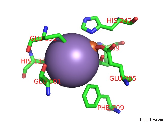
Mono view
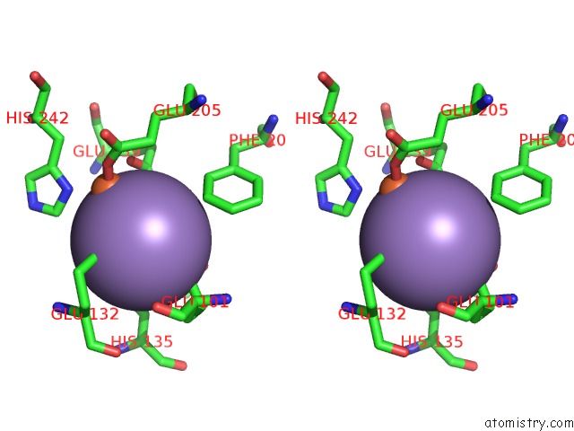
Stereo pair view
Mono view
Stereo pair view
A full contact list of Manganese with other atoms in the Mn binding
site number 1 of Crystal Structure of Ribonucleotide Reductase R2 Subunit Solved By Serial Synchrotron Crystallography within 5.0Å range:
|
Reference:
A.Shilova,
H.Lebrette,
O.Aurelius,
J.Nan,
M.Welin,
R.Kovacic,
S.Ghosh,
C.Safari,
R.J.Friel,
M.Milas,
Z.Matej,
M.Hogbom,
G.Branden,
M.Kloos,
R.L.Shoeman,
B.Doak,
T.Ursby,
M.Hakansson,
D.T.Logan,
U.Mueller.
Current Status and Future Opportunities For Serial Crystallography at Max IV Laboratory. J.Synchrotron Radiat. V. 27 1095 2020.
ISSN: ESSN 1600-5775
PubMed: 32876583
DOI: 10.1107/S1600577520008735
Page generated: Sun Oct 6 07:53:31 2024
ISSN: ESSN 1600-5775
PubMed: 32876583
DOI: 10.1107/S1600577520008735
Last articles
Zn in 9MJ5Zn in 9HNW
Zn in 9G0L
Zn in 9FNE
Zn in 9DZN
Zn in 9E0I
Zn in 9D32
Zn in 9DAK
Zn in 8ZXC
Zn in 8ZUF