Manganese »
PDB 6wj4-6z6r »
6x7u »
Manganese in PDB 6x7u: Crystal Structure of the Human Nudix Hydrolase NUDT16 in Complex with Fad
Enzymatic activity of Crystal Structure of the Human Nudix Hydrolase NUDT16 in Complex with Fad
All present enzymatic activity of Crystal Structure of the Human Nudix Hydrolase NUDT16 in Complex with Fad:
3.6.1.62; 3.6.1.64;
3.6.1.62; 3.6.1.64;
Protein crystallography data
The structure of Crystal Structure of the Human Nudix Hydrolase NUDT16 in Complex with Fad, PDB code: 6x7u
was solved by
K.Hamilton,
L.Tong,
with X-Ray Crystallography technique. A brief refinement statistics is given in the table below:
| Resolution Low / High (Å) | 45.47 / 2.70 |
| Space group | P 32 |
| Cell size a, b, c (Å), α, β, γ (°) | 64.721, 64.721, 77.751, 90.00, 90.00, 120.00 |
| R / Rfree (%) | 15.9 / 23.1 |
Manganese Binding Sites:
The binding sites of Manganese atom in the Crystal Structure of the Human Nudix Hydrolase NUDT16 in Complex with Fad
(pdb code 6x7u). This binding sites where shown within
5.0 Angstroms radius around Manganese atom.
In total 4 binding sites of Manganese where determined in the Crystal Structure of the Human Nudix Hydrolase NUDT16 in Complex with Fad, PDB code: 6x7u:
Jump to Manganese binding site number: 1; 2; 3; 4;
In total 4 binding sites of Manganese where determined in the Crystal Structure of the Human Nudix Hydrolase NUDT16 in Complex with Fad, PDB code: 6x7u:
Jump to Manganese binding site number: 1; 2; 3; 4;
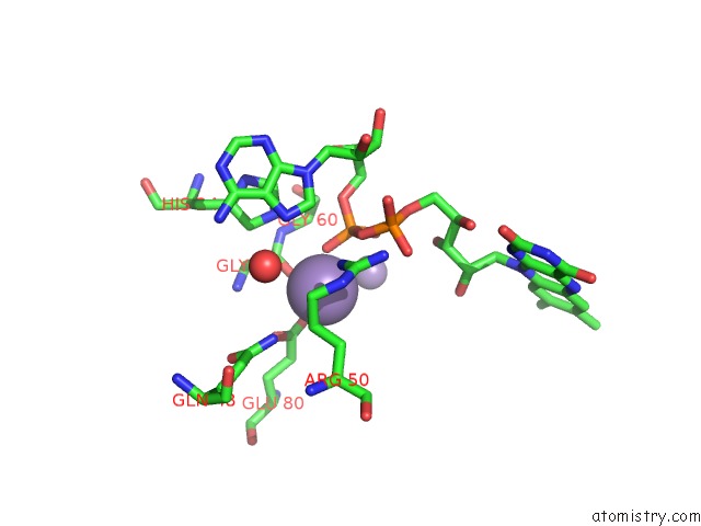
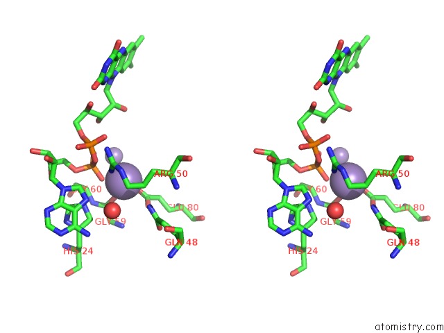
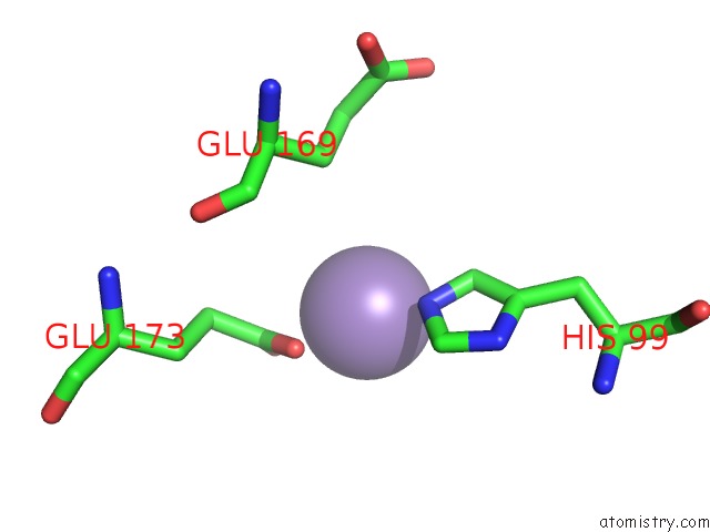
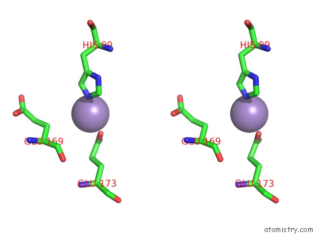
Manganese binding site 1 out of 4 in 6x7u
Go back to
Manganese binding site 1 out
of 4 in the Crystal Structure of the Human Nudix Hydrolase NUDT16 in Complex with Fad
Mono view
Stereo pair view
Mono view
Stereo pair view
A full contact list of Manganese with other atoms in the Mn binding
site number 1 of Crystal Structure of the Human Nudix Hydrolase NUDT16 in Complex with Fad within 5.0Å range:
|
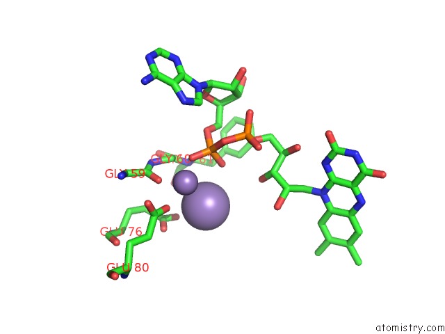
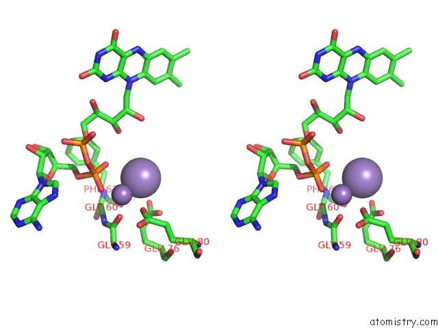
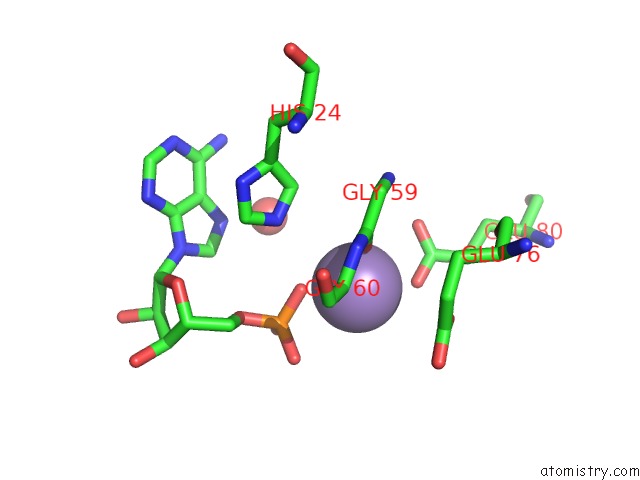
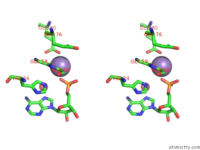
Manganese binding site 2 out of 4 in 6x7u
Go back to
Manganese binding site 2 out
of 4 in the Crystal Structure of the Human Nudix Hydrolase NUDT16 in Complex with Fad
Mono view
Stereo pair view
Mono view
Stereo pair view
A full contact list of Manganese with other atoms in the Mn binding
site number 2 of Crystal Structure of the Human Nudix Hydrolase NUDT16 in Complex with Fad within 5.0Å range:
|
Manganese binding site 3 out of 4 in 6x7u
Go back to
Manganese binding site 3 out
of 4 in the Crystal Structure of the Human Nudix Hydrolase NUDT16 in Complex with Fad
Mono view
Stereo pair view
Mono view
Stereo pair view
A full contact list of Manganese with other atoms in the Mn binding
site number 3 of Crystal Structure of the Human Nudix Hydrolase NUDT16 in Complex with Fad within 5.0Å range:
|
Manganese binding site 4 out of 4 in 6x7u
Go back to
Manganese binding site 4 out
of 4 in the Crystal Structure of the Human Nudix Hydrolase NUDT16 in Complex with Fad
Mono view
Stereo pair view
Mono view
Stereo pair view
A full contact list of Manganese with other atoms in the Mn binding
site number 4 of Crystal Structure of the Human Nudix Hydrolase NUDT16 in Complex with Fad within 5.0Å range:
|
Reference:
S.Sharma,
E.Grudzien-Nogalska,
K.Hamilton,
X.Jiao,
J.Yang,
L.Tong,
M.Kiledjian.
Mammalian Nudix Proteins Cleave Nucleotide Metabolite Caps on Rnas. Nucleic Acids Res. V. 48 6788 2020.
ISSN: ESSN 1362-4962
PubMed: 32432673
DOI: 10.1093/NAR/GKAA402
Page generated: Sun Oct 6 07:51:43 2024
ISSN: ESSN 1362-4962
PubMed: 32432673
DOI: 10.1093/NAR/GKAA402
Last articles
Zn in 9J0NZn in 9J0O
Zn in 9J0P
Zn in 9FJX
Zn in 9EKB
Zn in 9C0F
Zn in 9CAH
Zn in 9CH0
Zn in 9CH3
Zn in 9CH1