Manganese »
PDB 6wj4-6z6r »
6wn6 »
Manganese in PDB 6wn6: Crystal Structure of 3-Keto-D-Glucoside 4-Epimerase, Ycjr, From E. Coli, Apo Form
Protein crystallography data
The structure of Crystal Structure of 3-Keto-D-Glucoside 4-Epimerase, Ycjr, From E. Coli, Apo Form, PDB code: 6wn6
was solved by
M.F.Mabanglo,
F.M.Raushel,
K.Mukherjee,
with X-Ray Crystallography technique. A brief refinement statistics is given in the table below:
| Resolution Low / High (Å) | 35.91 / 1.86 |
| Space group | P 41 21 2 |
| Cell size a, b, c (Å), α, β, γ (°) | 116.576, 116.576, 247.725, 90.00, 90.00, 90.00 |
| R / Rfree (%) | 17.6 / 19.9 |
Manganese Binding Sites:
The binding sites of Manganese atom in the Crystal Structure of 3-Keto-D-Glucoside 4-Epimerase, Ycjr, From E. Coli, Apo Form
(pdb code 6wn6). This binding sites where shown within
5.0 Angstroms radius around Manganese atom.
In total 4 binding sites of Manganese where determined in the Crystal Structure of 3-Keto-D-Glucoside 4-Epimerase, Ycjr, From E. Coli, Apo Form, PDB code: 6wn6:
Jump to Manganese binding site number: 1; 2; 3; 4;
In total 4 binding sites of Manganese where determined in the Crystal Structure of 3-Keto-D-Glucoside 4-Epimerase, Ycjr, From E. Coli, Apo Form, PDB code: 6wn6:
Jump to Manganese binding site number: 1; 2; 3; 4;
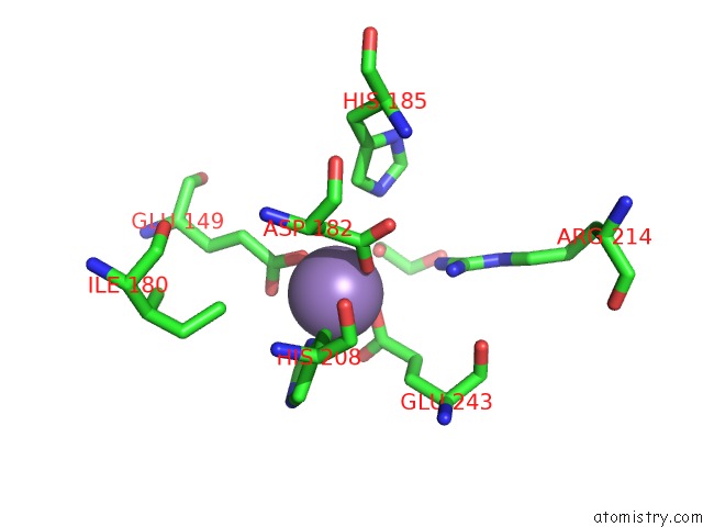
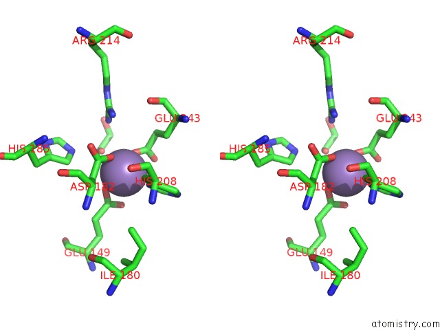
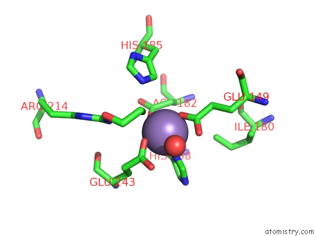
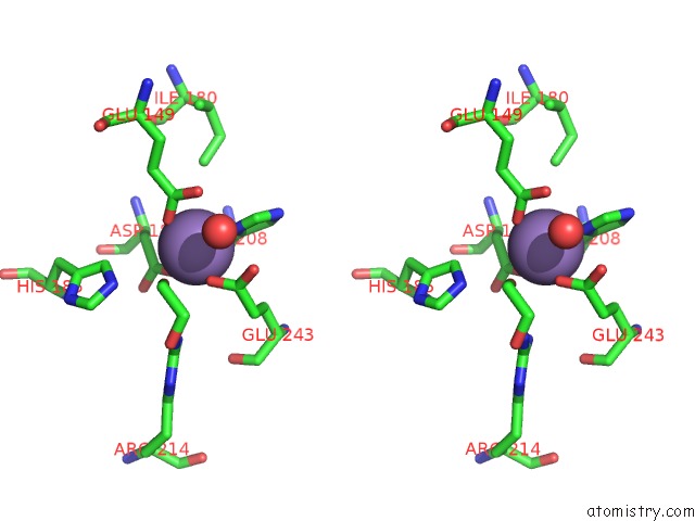
Manganese binding site 1 out of 4 in 6wn6
Go back to
Manganese binding site 1 out
of 4 in the Crystal Structure of 3-Keto-D-Glucoside 4-Epimerase, Ycjr, From E. Coli, Apo Form
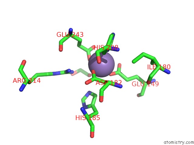
Mono view
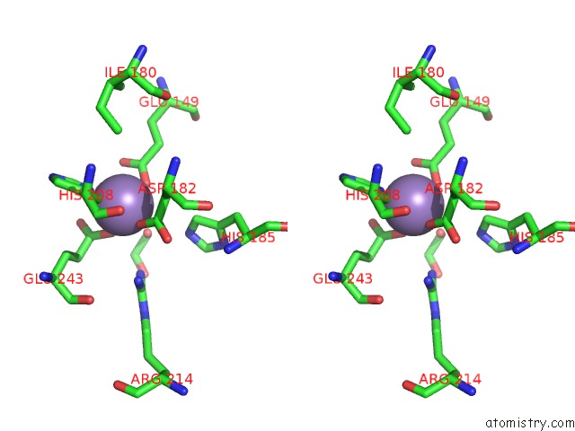
Stereo pair view
Mono view
Stereo pair view
A full contact list of Manganese with other atoms in the Mn binding
site number 1 of Crystal Structure of 3-Keto-D-Glucoside 4-Epimerase, Ycjr, From E. Coli, Apo Form within 5.0Å range:
|
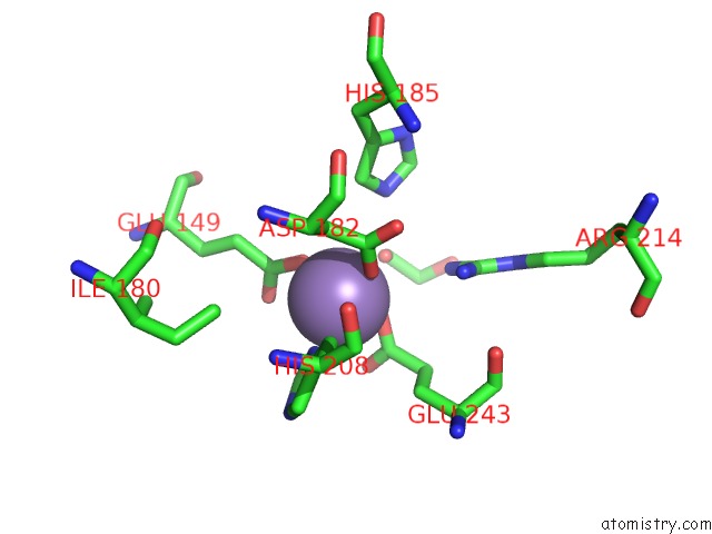
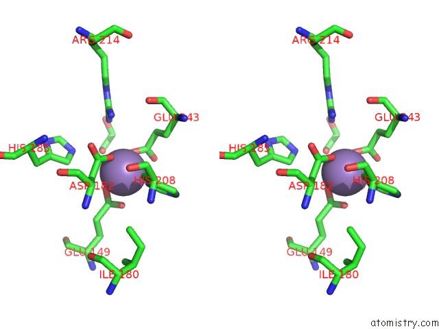
Manganese binding site 2 out of 4 in 6wn6
Go back to
Manganese binding site 2 out
of 4 in the Crystal Structure of 3-Keto-D-Glucoside 4-Epimerase, Ycjr, From E. Coli, Apo Form
Mono view
Stereo pair view
Mono view
Stereo pair view
A full contact list of Manganese with other atoms in the Mn binding
site number 2 of Crystal Structure of 3-Keto-D-Glucoside 4-Epimerase, Ycjr, From E. Coli, Apo Form within 5.0Å range:
|
Manganese binding site 3 out of 4 in 6wn6
Go back to
Manganese binding site 3 out
of 4 in the Crystal Structure of 3-Keto-D-Glucoside 4-Epimerase, Ycjr, From E. Coli, Apo Form
Mono view
Stereo pair view
Mono view
Stereo pair view
A full contact list of Manganese with other atoms in the Mn binding
site number 3 of Crystal Structure of 3-Keto-D-Glucoside 4-Epimerase, Ycjr, From E. Coli, Apo Form within 5.0Å range:
|
Manganese binding site 4 out of 4 in 6wn6
Go back to
Manganese binding site 4 out
of 4 in the Crystal Structure of 3-Keto-D-Glucoside 4-Epimerase, Ycjr, From E. Coli, Apo Form
Mono view
Stereo pair view
Mono view
Stereo pair view
A full contact list of Manganese with other atoms in the Mn binding
site number 4 of Crystal Structure of 3-Keto-D-Glucoside 4-Epimerase, Ycjr, From E. Coli, Apo Form within 5.0Å range:
|
Reference:
M.F.Mabanglo,
J.P.Huddleston,
K.Mukherjee,
Z.W.Taylor,
F.M.Raushel.
Structure and Reaction Mechanism of Ycjr, An Epimerase That Facilitates the Interconversion of D-Gulosides to D-Glucosides Inescherichia Coli. Biochemistry 2020.
ISSN: ISSN 0006-2960
PubMed: 32437133
DOI: 10.1021/ACS.BIOCHEM.0C00334
Page generated: Sun Oct 6 07:51:04 2024
ISSN: ISSN 0006-2960
PubMed: 32437133
DOI: 10.1021/ACS.BIOCHEM.0C00334
Last articles
Zn in 9J0NZn in 9J0O
Zn in 9J0P
Zn in 9FJX
Zn in 9EKB
Zn in 9C0F
Zn in 9CAH
Zn in 9CH0
Zn in 9CH3
Zn in 9CH1