Manganese »
PDB 6vf5-6wij »
6wbt »
Manganese in PDB 6wbt: 2.52 Angstrom Resolution Crystal Structure of 6-Phospho-Alpha- Glucosidase From Gut Microorganisms in Complex with Nad and Glucose- 6-Phosphate
Protein crystallography data
The structure of 2.52 Angstrom Resolution Crystal Structure of 6-Phospho-Alpha- Glucosidase From Gut Microorganisms in Complex with Nad and Glucose- 6-Phosphate, PDB code: 6wbt
was solved by
R.Wu,
Y.Kim,
M.Endres,
J.Joachimiak,
with X-Ray Crystallography technique. A brief refinement statistics is given in the table below:
| Resolution Low / High (Å) | 70.92 / 2.52 |
| Space group | P 21 21 21 |
| Cell size a, b, c (Å), α, β, γ (°) | 102.797, 219.724, 97.978, 90, 90, 90 |
| R / Rfree (%) | 22.7 / 26.6 |
Manganese Binding Sites:
The binding sites of Manganese atom in the 2.52 Angstrom Resolution Crystal Structure of 6-Phospho-Alpha- Glucosidase From Gut Microorganisms in Complex with Nad and Glucose- 6-Phosphate
(pdb code 6wbt). This binding sites where shown within
5.0 Angstroms radius around Manganese atom.
In total 4 binding sites of Manganese where determined in the 2.52 Angstrom Resolution Crystal Structure of 6-Phospho-Alpha- Glucosidase From Gut Microorganisms in Complex with Nad and Glucose- 6-Phosphate, PDB code: 6wbt:
Jump to Manganese binding site number: 1; 2; 3; 4;
In total 4 binding sites of Manganese where determined in the 2.52 Angstrom Resolution Crystal Structure of 6-Phospho-Alpha- Glucosidase From Gut Microorganisms in Complex with Nad and Glucose- 6-Phosphate, PDB code: 6wbt:
Jump to Manganese binding site number: 1; 2; 3; 4;
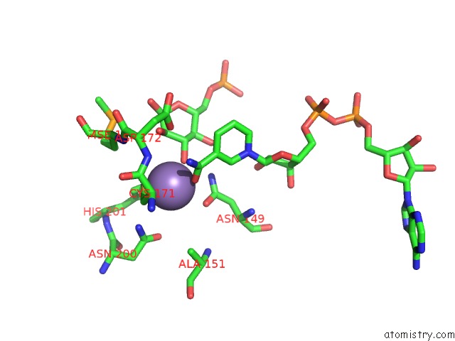
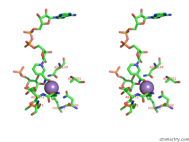
Manganese binding site 1 out of 4 in 6wbt
Go back to
Manganese binding site 1 out
of 4 in the 2.52 Angstrom Resolution Crystal Structure of 6-Phospho-Alpha- Glucosidase From Gut Microorganisms in Complex with Nad and Glucose- 6-Phosphate
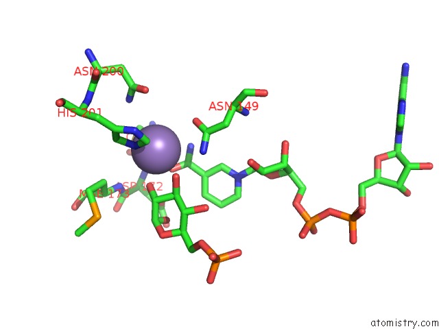
Mono view
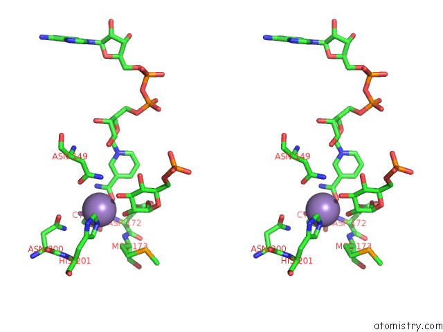
Stereo pair view
Mono view
Stereo pair view
A full contact list of Manganese with other atoms in the Mn binding
site number 1 of 2.52 Angstrom Resolution Crystal Structure of 6-Phospho-Alpha- Glucosidase From Gut Microorganisms in Complex with Nad and Glucose- 6-Phosphate within 5.0Å range:
|
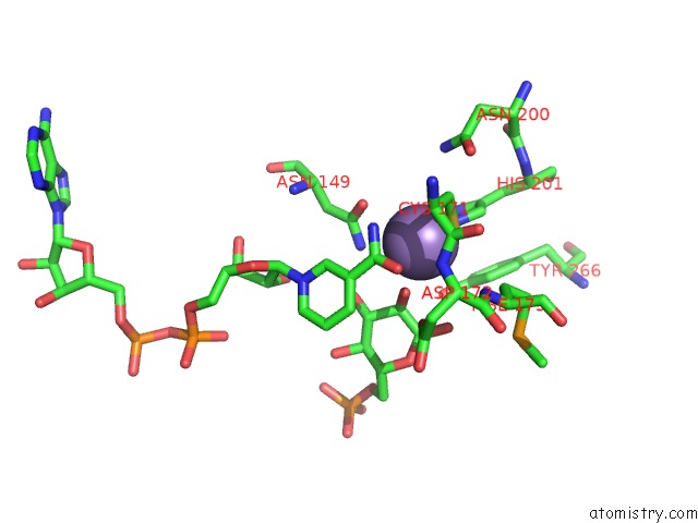
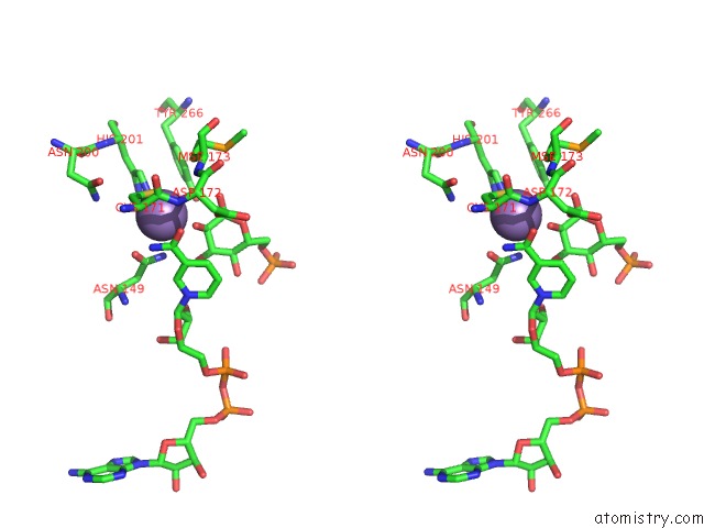
Manganese binding site 2 out of 4 in 6wbt
Go back to
Manganese binding site 2 out
of 4 in the 2.52 Angstrom Resolution Crystal Structure of 6-Phospho-Alpha- Glucosidase From Gut Microorganisms in Complex with Nad and Glucose- 6-Phosphate
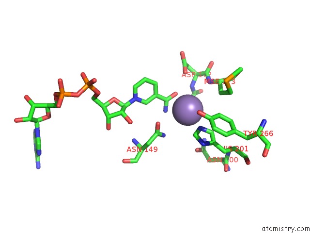
Mono view
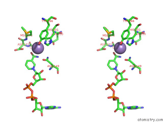
Stereo pair view
Mono view
Stereo pair view
A full contact list of Manganese with other atoms in the Mn binding
site number 2 of 2.52 Angstrom Resolution Crystal Structure of 6-Phospho-Alpha- Glucosidase From Gut Microorganisms in Complex with Nad and Glucose- 6-Phosphate within 5.0Å range:
|
Manganese binding site 3 out of 4 in 6wbt
Go back to
Manganese binding site 3 out
of 4 in the 2.52 Angstrom Resolution Crystal Structure of 6-Phospho-Alpha- Glucosidase From Gut Microorganisms in Complex with Nad and Glucose- 6-Phosphate
Mono view
Stereo pair view
Mono view
Stereo pair view
A full contact list of Manganese with other atoms in the Mn binding
site number 3 of 2.52 Angstrom Resolution Crystal Structure of 6-Phospho-Alpha- Glucosidase From Gut Microorganisms in Complex with Nad and Glucose- 6-Phosphate within 5.0Å range:
|
Manganese binding site 4 out of 4 in 6wbt
Go back to
Manganese binding site 4 out
of 4 in the 2.52 Angstrom Resolution Crystal Structure of 6-Phospho-Alpha- Glucosidase From Gut Microorganisms in Complex with Nad and Glucose- 6-Phosphate
Mono view
Stereo pair view
Mono view
Stereo pair view
A full contact list of Manganese with other atoms in the Mn binding
site number 4 of 2.52 Angstrom Resolution Crystal Structure of 6-Phospho-Alpha- Glucosidase From Gut Microorganisms in Complex with Nad and Glucose- 6-Phosphate within 5.0Å range:
|
Reference:
R.Wu,
Y.Kim,
M.Endres,
J.Joachimiak.
2.52 Angstrom Resolution Crystal Structure of 6-Phospho-Alpha-Glucosidase From Gut Microorganisms in Complex with Nad and Glucose-6-Phosphate To Be Published.
Page generated: Sun Oct 6 07:42:19 2024
Last articles
Cl in 3CCMCl in 3CCL
Cl in 3CCJ
Cl in 3CCE
Cl in 3CCK
Cl in 3CC4
Cl in 3CC7
Cl in 3CC2
Cl in 3CCC
Cl in 3C8V