Manganese »
PDB 6tzp-6vf4 »
6vdx »
Manganese in PDB 6vdx: Crystal Structure of Dehaloperoxidase B in Complex with Cofactor Manganese(III)- Porphyrin and Substrate Trichlorophenol
Protein crystallography data
The structure of Crystal Structure of Dehaloperoxidase B in Complex with Cofactor Manganese(III)- Porphyrin and Substrate Trichlorophenol, PDB code: 6vdx
was solved by
R.A.Ghiladi,
V.S.De Serrano,
A.Mcguire,
T.Malewschik,
with X-Ray Crystallography technique. A brief refinement statistics is given in the table below:
| Resolution Low / High (Å) | 44.64 / 1.53 |
| Space group | P 21 21 21 |
| Cell size a, b, c (Å), α, β, γ (°) | 59.365, 67.585, 67.626, 90, 90, 90 |
| R / Rfree (%) | 16.5 / 23.7 |
Other elements in 6vdx:
The structure of Crystal Structure of Dehaloperoxidase B in Complex with Cofactor Manganese(III)- Porphyrin and Substrate Trichlorophenol also contains other interesting chemical elements:
| Chlorine | (Cl) | 6 atoms |
Manganese Binding Sites:
The binding sites of Manganese atom in the Crystal Structure of Dehaloperoxidase B in Complex with Cofactor Manganese(III)- Porphyrin and Substrate Trichlorophenol
(pdb code 6vdx). This binding sites where shown within
5.0 Angstroms radius around Manganese atom.
In total 2 binding sites of Manganese where determined in the Crystal Structure of Dehaloperoxidase B in Complex with Cofactor Manganese(III)- Porphyrin and Substrate Trichlorophenol, PDB code: 6vdx:
Jump to Manganese binding site number: 1; 2;
In total 2 binding sites of Manganese where determined in the Crystal Structure of Dehaloperoxidase B in Complex with Cofactor Manganese(III)- Porphyrin and Substrate Trichlorophenol, PDB code: 6vdx:
Jump to Manganese binding site number: 1; 2;
Manganese binding site 1 out of 2 in 6vdx
Go back to
Manganese binding site 1 out
of 2 in the Crystal Structure of Dehaloperoxidase B in Complex with Cofactor Manganese(III)- Porphyrin and Substrate Trichlorophenol
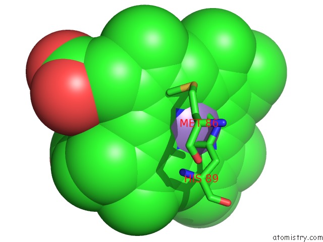
Mono view
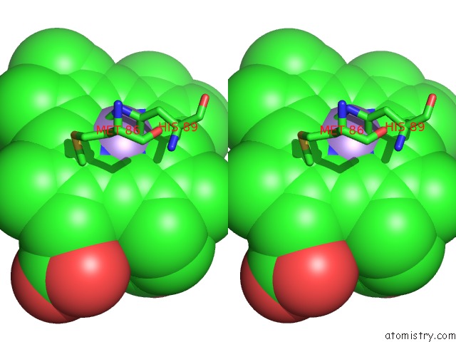
Stereo pair view
Mono view
Stereo pair view
A full contact list of Manganese with other atoms in the Mn binding
site number 1 of Crystal Structure of Dehaloperoxidase B in Complex with Cofactor Manganese(III)- Porphyrin and Substrate Trichlorophenol within 5.0Å range:
|
Manganese binding site 2 out of 2 in 6vdx
Go back to
Manganese binding site 2 out
of 2 in the Crystal Structure of Dehaloperoxidase B in Complex with Cofactor Manganese(III)- Porphyrin and Substrate Trichlorophenol
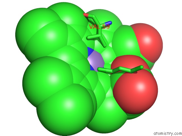
Mono view
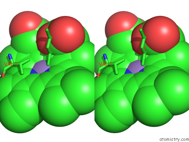
Stereo pair view
Mono view
Stereo pair view
A full contact list of Manganese with other atoms in the Mn binding
site number 2 of Crystal Structure of Dehaloperoxidase B in Complex with Cofactor Manganese(III)- Porphyrin and Substrate Trichlorophenol within 5.0Å range:
|
Reference:
A.H.Mcguire,
A.R.Petit,
J.Kang,
T.Malewschik,
V.De Serrano,
L.M.Carey,
R.A.Ghiladi.
Nonnative Heme Incorporation Into Multifunctional Globin Increases Peroxygenase Activity An Order and Magnitude Compared to Native Enzyme To Be Published.
Page generated: Sun Oct 6 07:29:21 2024
Last articles
Zn in 9J0NZn in 9J0O
Zn in 9J0P
Zn in 9FJX
Zn in 9EKB
Zn in 9C0F
Zn in 9CAH
Zn in 9CH0
Zn in 9CH3
Zn in 9CH1