Manganese »
PDB 6tzp-6vf4 »
6vc6 »
Manganese in PDB 6vc6: 2.1 Angstrom Resolution Crystal Structure of 6-Phospho-Alpha- Glucosidase From Gut Microorganisms in Complex with Nad and MN2+
Enzymatic activity of 2.1 Angstrom Resolution Crystal Structure of 6-Phospho-Alpha- Glucosidase From Gut Microorganisms in Complex with Nad and MN2+
All present enzymatic activity of 2.1 Angstrom Resolution Crystal Structure of 6-Phospho-Alpha- Glucosidase From Gut Microorganisms in Complex with Nad and MN2+:
3.2.1.122;
3.2.1.122;
Protein crystallography data
The structure of 2.1 Angstrom Resolution Crystal Structure of 6-Phospho-Alpha- Glucosidase From Gut Microorganisms in Complex with Nad and MN2+, PDB code: 6vc6
was solved by
R.Wu,
Y.Kim,
M.Endres,
J.Joachimiak,
with X-Ray Crystallography technique. A brief refinement statistics is given in the table below:
| Resolution Low / High (Å) | 48.61 / 2.13 |
| Space group | C 1 2 1 |
| Cell size a, b, c (Å), α, β, γ (°) | 194.460, 62.257, 160.706, 90.00, 90.35, 90.00 |
| R / Rfree (%) | 19.6 / 22.9 |
Manganese Binding Sites:
The binding sites of Manganese atom in the 2.1 Angstrom Resolution Crystal Structure of 6-Phospho-Alpha- Glucosidase From Gut Microorganisms in Complex with Nad and MN2+
(pdb code 6vc6). This binding sites where shown within
5.0 Angstroms radius around Manganese atom.
In total 4 binding sites of Manganese where determined in the 2.1 Angstrom Resolution Crystal Structure of 6-Phospho-Alpha- Glucosidase From Gut Microorganisms in Complex with Nad and MN2+, PDB code: 6vc6:
Jump to Manganese binding site number: 1; 2; 3; 4;
In total 4 binding sites of Manganese where determined in the 2.1 Angstrom Resolution Crystal Structure of 6-Phospho-Alpha- Glucosidase From Gut Microorganisms in Complex with Nad and MN2+, PDB code: 6vc6:
Jump to Manganese binding site number: 1; 2; 3; 4;
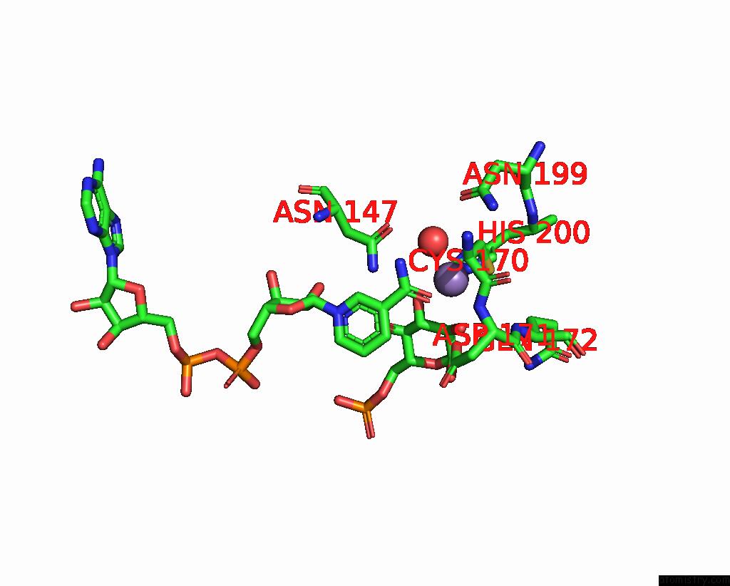
Manganese binding site 1 out of 4 in 6vc6
Go back to
Manganese binding site 1 out
of 4 in the 2.1 Angstrom Resolution Crystal Structure of 6-Phospho-Alpha- Glucosidase From Gut Microorganisms in Complex with Nad and MN2+
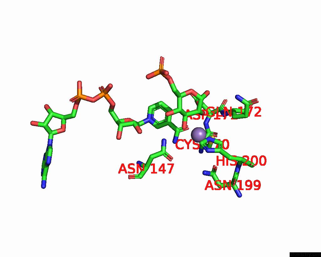
Mono view
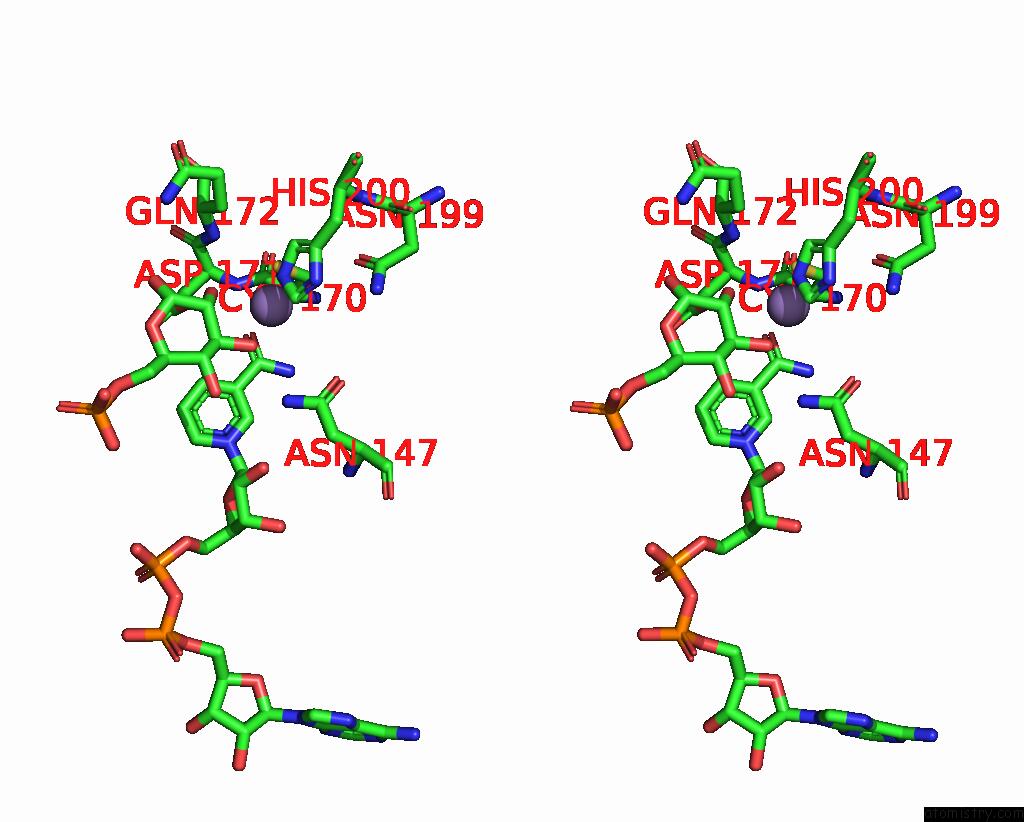
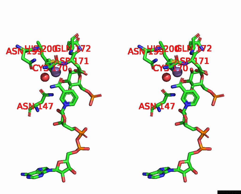
Stereo pair view
Mono view
Stereo pair view
A full contact list of Manganese with other atoms in the Mn binding
site number 1 of 2.1 Angstrom Resolution Crystal Structure of 6-Phospho-Alpha- Glucosidase From Gut Microorganisms in Complex with Nad and MN2+ within 5.0Å range:
|
Manganese binding site 2 out of 4 in 6vc6
Go back to
Manganese binding site 2 out
of 4 in the 2.1 Angstrom Resolution Crystal Structure of 6-Phospho-Alpha- Glucosidase From Gut Microorganisms in Complex with Nad and MN2+
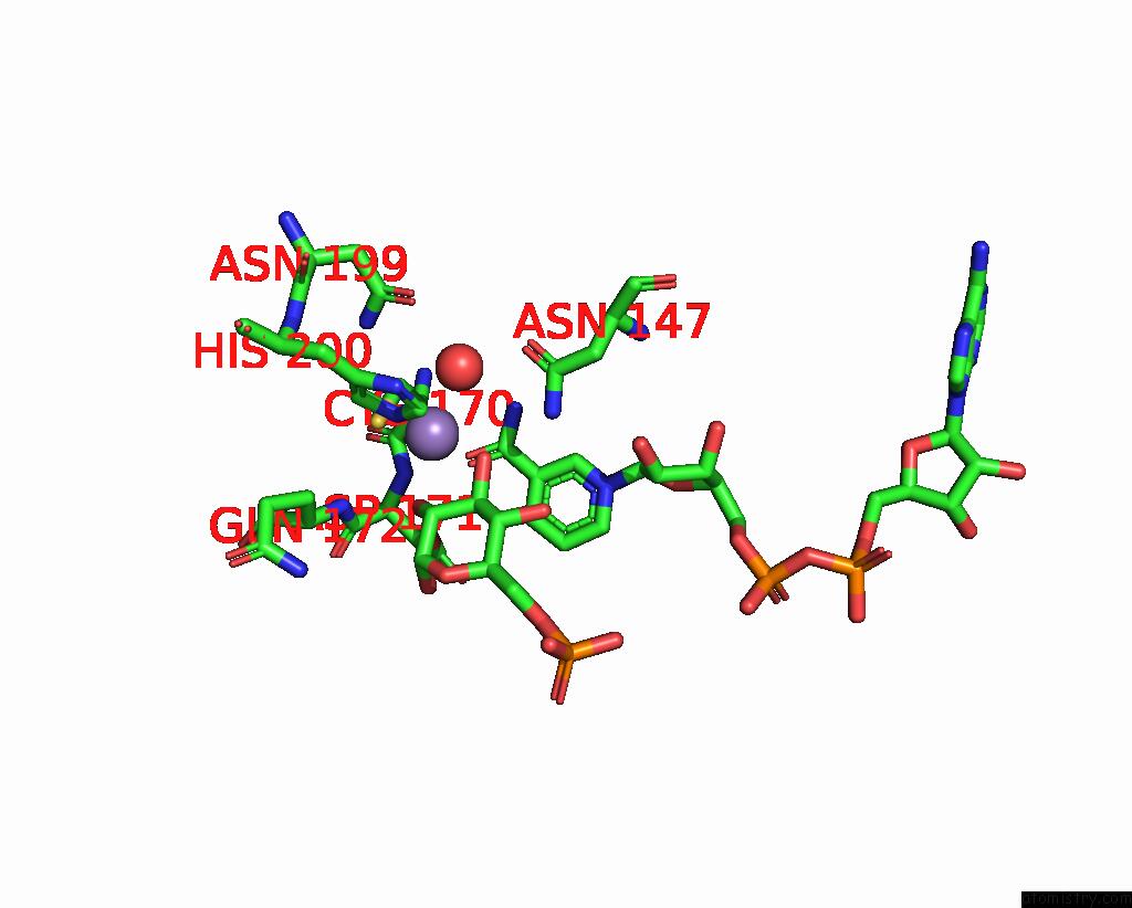
Mono view
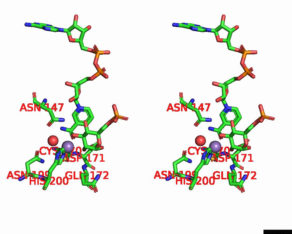
Stereo pair view
Mono view
Stereo pair view
A full contact list of Manganese with other atoms in the Mn binding
site number 2 of 2.1 Angstrom Resolution Crystal Structure of 6-Phospho-Alpha- Glucosidase From Gut Microorganisms in Complex with Nad and MN2+ within 5.0Å range:
|
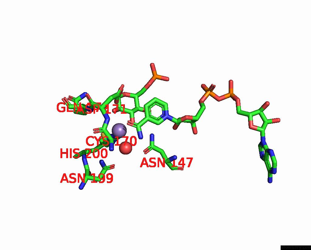
Manganese binding site 3 out of 4 in 6vc6
Go back to
Manganese binding site 3 out
of 4 in the 2.1 Angstrom Resolution Crystal Structure of 6-Phospho-Alpha- Glucosidase From Gut Microorganisms in Complex with Nad and MN2+
Mono view
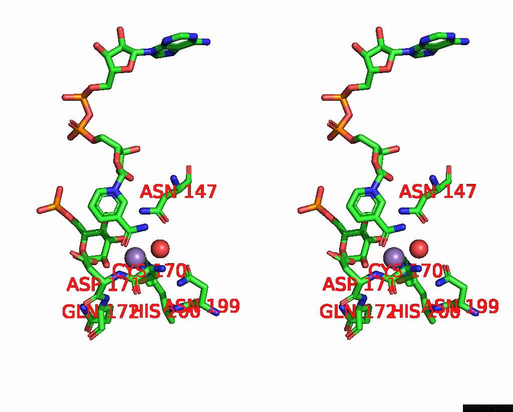
Stereo pair view
Mono view
Stereo pair view
A full contact list of Manganese with other atoms in the Mn binding
site number 3 of 2.1 Angstrom Resolution Crystal Structure of 6-Phospho-Alpha- Glucosidase From Gut Microorganisms in Complex with Nad and MN2+ within 5.0Å range:
|
Manganese binding site 4 out of 4 in 6vc6
Go back to
Manganese binding site 4 out
of 4 in the 2.1 Angstrom Resolution Crystal Structure of 6-Phospho-Alpha- Glucosidase From Gut Microorganisms in Complex with Nad and MN2+
Mono view
Stereo pair view
Mono view
Stereo pair view
A full contact list of Manganese with other atoms in the Mn binding
site number 4 of 2.1 Angstrom Resolution Crystal Structure of 6-Phospho-Alpha- Glucosidase From Gut Microorganisms in Complex with Nad and MN2+ within 5.0Å range:
|
Reference:
R.Wu,
Y.Kim,
M.Endres,
A.Joachimiak.
2.1 Angstrom Resolution Crystal Structure of 6-Phospho-Alpha-Glucosidase From Gut Microorganisms in Complex with Nad and MN2+ To Be Published.
Page generated: Sat Aug 16 21:51:48 2025
Last articles
Mo in 3NS1Mo in 3MIN
Mo in 3NRZ
Mo in 3K7R
Mo in 3ML1
Mo in 3K6X
Mo in 3L4P
Mo in 3K1A
Mo in 3K6W
Mo in 3GZG