Manganese »
PDB 6tzp-6vf4 »
6vaw »
Manganese in PDB 6vaw: Peanut Lectin Complexed with N-Beta-D-Galactopyranosyl-L-Succinamoyl Derivative (Ngs)
Protein crystallography data
The structure of Peanut Lectin Complexed with N-Beta-D-Galactopyranosyl-L-Succinamoyl Derivative (Ngs), PDB code: 6vaw
was solved by
L.H.Otero,
E.D.Primo,
A.J.Cagnoni,
S.Klinke,
F.A.Goldbaum,
M.L.Uhrig,
with X-Ray Crystallography technique. A brief refinement statistics is given in the table below:
| Resolution Low / High (Å) | 45.42 / 1.75 |
| Space group | P 2 21 21 |
| Cell size a, b, c (Å), α, β, γ (°) | 76.222, 125.052, 126.844, 90.00, 90.00, 90.00 |
| R / Rfree (%) | 21.2 / 23.2 |
Other elements in 6vaw:
The structure of Peanut Lectin Complexed with N-Beta-D-Galactopyranosyl-L-Succinamoyl Derivative (Ngs) also contains other interesting chemical elements:
| Calcium | (Ca) | 4 atoms |
Manganese Binding Sites:
The binding sites of Manganese atom in the Peanut Lectin Complexed with N-Beta-D-Galactopyranosyl-L-Succinamoyl Derivative (Ngs)
(pdb code 6vaw). This binding sites where shown within
5.0 Angstroms radius around Manganese atom.
In total 4 binding sites of Manganese where determined in the Peanut Lectin Complexed with N-Beta-D-Galactopyranosyl-L-Succinamoyl Derivative (Ngs), PDB code: 6vaw:
Jump to Manganese binding site number: 1; 2; 3; 4;
In total 4 binding sites of Manganese where determined in the Peanut Lectin Complexed with N-Beta-D-Galactopyranosyl-L-Succinamoyl Derivative (Ngs), PDB code: 6vaw:
Jump to Manganese binding site number: 1; 2; 3; 4;
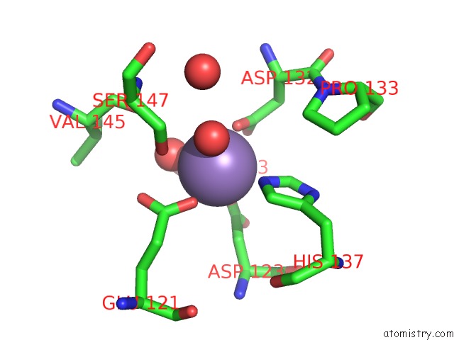
Manganese binding site 1 out of 4 in 6vaw
Go back to
Manganese binding site 1 out
of 4 in the Peanut Lectin Complexed with N-Beta-D-Galactopyranosyl-L-Succinamoyl Derivative (Ngs)
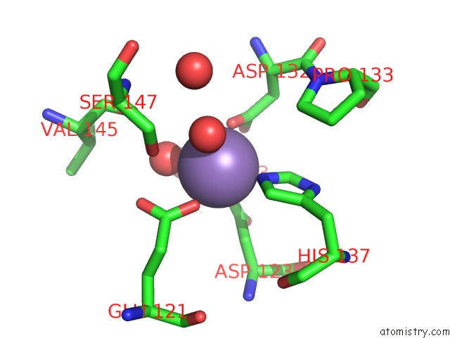
Mono view
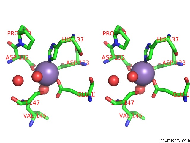
Stereo pair view
Mono view
Stereo pair view
A full contact list of Manganese with other atoms in the Mn binding
site number 1 of Peanut Lectin Complexed with N-Beta-D-Galactopyranosyl-L-Succinamoyl Derivative (Ngs) within 5.0Å range:
|
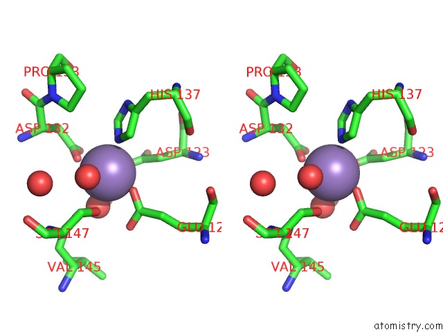
Manganese binding site 2 out of 4 in 6vaw
Go back to
Manganese binding site 2 out
of 4 in the Peanut Lectin Complexed with N-Beta-D-Galactopyranosyl-L-Succinamoyl Derivative (Ngs)
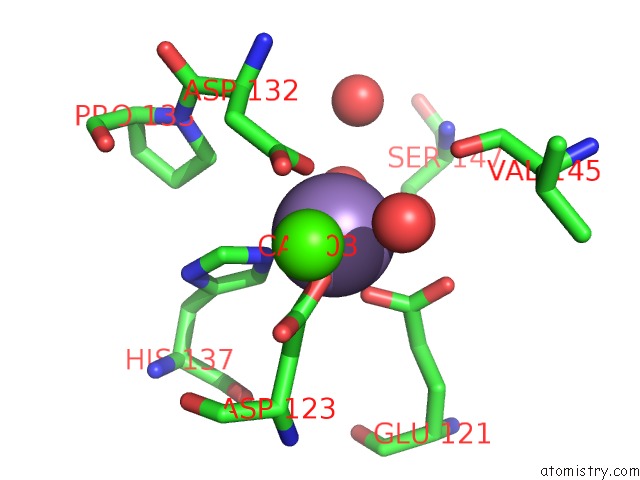
Mono view
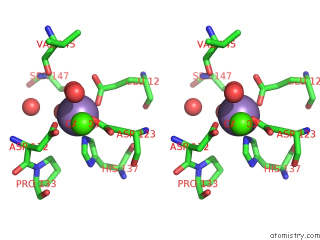
Stereo pair view
Mono view
Stereo pair view
A full contact list of Manganese with other atoms in the Mn binding
site number 2 of Peanut Lectin Complexed with N-Beta-D-Galactopyranosyl-L-Succinamoyl Derivative (Ngs) within 5.0Å range:
|
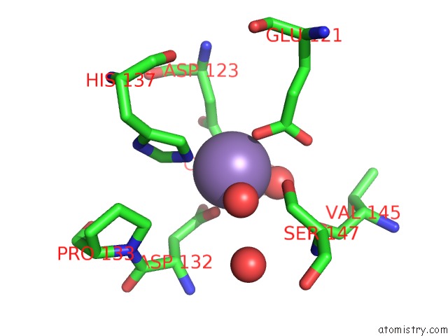
Manganese binding site 3 out of 4 in 6vaw
Go back to
Manganese binding site 3 out
of 4 in the Peanut Lectin Complexed with N-Beta-D-Galactopyranosyl-L-Succinamoyl Derivative (Ngs)
Mono view
Stereo pair view
Mono view
Stereo pair view
A full contact list of Manganese with other atoms in the Mn binding
site number 3 of Peanut Lectin Complexed with N-Beta-D-Galactopyranosyl-L-Succinamoyl Derivative (Ngs) within 5.0Å range:
|
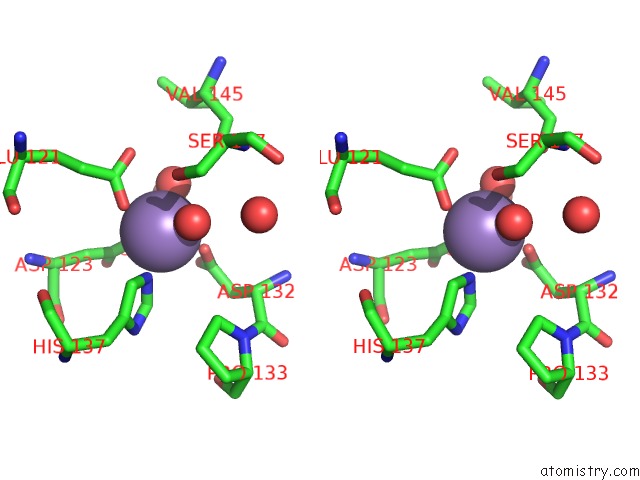
Manganese binding site 4 out of 4 in 6vaw
Go back to
Manganese binding site 4 out
of 4 in the Peanut Lectin Complexed with N-Beta-D-Galactopyranosyl-L-Succinamoyl Derivative (Ngs)
Mono view
Stereo pair view
Mono view
Stereo pair view
A full contact list of Manganese with other atoms in the Mn binding
site number 4 of Peanut Lectin Complexed with N-Beta-D-Galactopyranosyl-L-Succinamoyl Derivative (Ngs) within 5.0Å range:
|
Reference:
A.J.Cagnoni,
E.D.Primo,
S.Klinke,
M.E.Cano,
W.Giordano,
K.V.Marino,
J.Kovensky,
F.A.Goldbaum,
M.L.Uhrig,
L.H.Otero.
Crystal Structures of Peanut Lectin in the Presence of Synthetic Beta-N- and Beta-S-Galactosides Disclose Evidence For the Recognition of Different Glycomimetic Ligands. Acta Crystallogr D Struct V. 76 1080 2020BIOL.
ISSN: ISSN 2059-7983
PubMed: 33135679
DOI: 10.1107/S2059798320012371
Page generated: Sun Oct 6 07:25:38 2024
ISSN: ISSN 2059-7983
PubMed: 33135679
DOI: 10.1107/S2059798320012371
Last articles
Ca in 5UVLCa in 5UVG
Ca in 5UUE
Ca in 5UUY
Ca in 5UUH
Ca in 5UUG
Ca in 5UUD
Ca in 5UUC
Ca in 5UUB
Ca in 5UUA