Manganese »
PDB 6rwz-6txf »
6tfh »
Manganese in PDB 6tfh: Crystal Structure of the Adp-Binding Domain of the Nad+ Riboswitch with Nicotinamide Adenine Dinucleotide, Reduced (Nadh); Soaking with Manganese(II) (MN2+)
Protein crystallography data
The structure of Crystal Structure of the Adp-Binding Domain of the Nad+ Riboswitch with Nicotinamide Adenine Dinucleotide, Reduced (Nadh); Soaking with Manganese(II) (MN2+), PDB code: 6tfh
was solved by
L.Huang,
D.M.J.Lilley,
with X-Ray Crystallography technique. A brief refinement statistics is given in the table below:
| Resolution Low / High (Å) | 48.49 / 2.95 |
| Space group | I 2 2 2 |
| Cell size a, b, c (Å), α, β, γ (°) | 56.935, 58.360, 193.968, 90.00, 90.00, 90.00 |
| R / Rfree (%) | 22.1 / 26.6 |
Other elements in 6tfh:
The structure of Crystal Structure of the Adp-Binding Domain of the Nad+ Riboswitch with Nicotinamide Adenine Dinucleotide, Reduced (Nadh); Soaking with Manganese(II) (MN2+) also contains other interesting chemical elements:
| Bromine | (Br) | 1 atom |
| Sodium | (Na) | 1 atom |
Manganese Binding Sites:
The binding sites of Manganese atom in the Crystal Structure of the Adp-Binding Domain of the Nad+ Riboswitch with Nicotinamide Adenine Dinucleotide, Reduced (Nadh); Soaking with Manganese(II) (MN2+)
(pdb code 6tfh). This binding sites where shown within
5.0 Angstroms radius around Manganese atom.
In total 9 binding sites of Manganese where determined in the Crystal Structure of the Adp-Binding Domain of the Nad+ Riboswitch with Nicotinamide Adenine Dinucleotide, Reduced (Nadh); Soaking with Manganese(II) (MN2+), PDB code: 6tfh:
Jump to Manganese binding site number: 1; 2; 3; 4; 5; 6; 7; 8; 9;
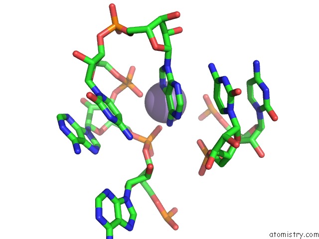
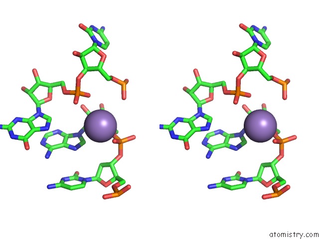
Manganese binding site 1 out of 9 in 6tfh
Go back to
Manganese binding site 1 out
of 9 in the Crystal Structure of the Adp-Binding Domain of the Nad+ Riboswitch with Nicotinamide Adenine Dinucleotide, Reduced (Nadh); Soaking with Manganese(II) (MN2+)

Mono view

Stereo pair view
Mono view
Stereo pair view
A full contact list of Manganese with other atoms in the Mn binding
site number 1 of Crystal Structure of the Adp-Binding Domain of the Nad+ Riboswitch with Nicotinamide Adenine Dinucleotide, Reduced (Nadh); Soaking with Manganese(II) (MN2+) within 5.0Å range:
|
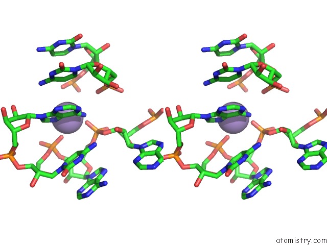
Manganese binding site 2 out of 9 in 6tfh
Go back to
Manganese binding site 2 out
of 9 in the Crystal Structure of the Adp-Binding Domain of the Nad+ Riboswitch with Nicotinamide Adenine Dinucleotide, Reduced (Nadh); Soaking with Manganese(II) (MN2+)

Mono view

Stereo pair view
Mono view
Stereo pair view
A full contact list of Manganese with other atoms in the Mn binding
site number 2 of Crystal Structure of the Adp-Binding Domain of the Nad+ Riboswitch with Nicotinamide Adenine Dinucleotide, Reduced (Nadh); Soaking with Manganese(II) (MN2+) within 5.0Å range:
|
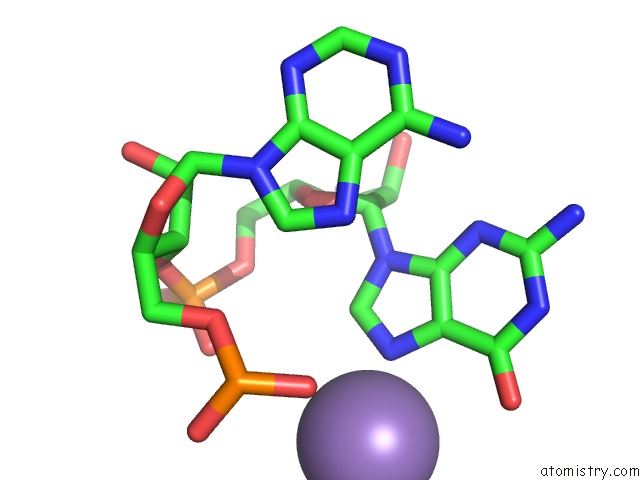
Manganese binding site 3 out of 9 in 6tfh
Go back to
Manganese binding site 3 out
of 9 in the Crystal Structure of the Adp-Binding Domain of the Nad+ Riboswitch with Nicotinamide Adenine Dinucleotide, Reduced (Nadh); Soaking with Manganese(II) (MN2+)

Mono view

Stereo pair view
Mono view
Stereo pair view
A full contact list of Manganese with other atoms in the Mn binding
site number 3 of Crystal Structure of the Adp-Binding Domain of the Nad+ Riboswitch with Nicotinamide Adenine Dinucleotide, Reduced (Nadh); Soaking with Manganese(II) (MN2+) within 5.0Å range:
|
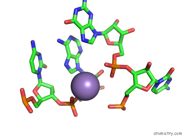
Manganese binding site 4 out of 9 in 6tfh
Go back to
Manganese binding site 4 out
of 9 in the Crystal Structure of the Adp-Binding Domain of the Nad+ Riboswitch with Nicotinamide Adenine Dinucleotide, Reduced (Nadh); Soaking with Manganese(II) (MN2+)
Mono view
Stereo pair view
Mono view
Stereo pair view
A full contact list of Manganese with other atoms in the Mn binding
site number 4 of Crystal Structure of the Adp-Binding Domain of the Nad+ Riboswitch with Nicotinamide Adenine Dinucleotide, Reduced (Nadh); Soaking with Manganese(II) (MN2+) within 5.0Å range:
|
Manganese binding site 5 out of 9 in 6tfh
Go back to
Manganese binding site 5 out
of 9 in the Crystal Structure of the Adp-Binding Domain of the Nad+ Riboswitch with Nicotinamide Adenine Dinucleotide, Reduced (Nadh); Soaking with Manganese(II) (MN2+)
Mono view

Stereo pair view
Mono view
Stereo pair view
A full contact list of Manganese with other atoms in the Mn binding
site number 5 of Crystal Structure of the Adp-Binding Domain of the Nad+ Riboswitch with Nicotinamide Adenine Dinucleotide, Reduced (Nadh); Soaking with Manganese(II) (MN2+) within 5.0Å range:
|
Manganese binding site 6 out of 9 in 6tfh
Go back to
Manganese binding site 6 out
of 9 in the Crystal Structure of the Adp-Binding Domain of the Nad+ Riboswitch with Nicotinamide Adenine Dinucleotide, Reduced (Nadh); Soaking with Manganese(II) (MN2+)

Mono view

Stereo pair view
Mono view
Stereo pair view
A full contact list of Manganese with other atoms in the Mn binding
site number 6 of Crystal Structure of the Adp-Binding Domain of the Nad+ Riboswitch with Nicotinamide Adenine Dinucleotide, Reduced (Nadh); Soaking with Manganese(II) (MN2+) within 5.0Å range:
|
Manganese binding site 7 out of 9 in 6tfh
Go back to
Manganese binding site 7 out
of 9 in the Crystal Structure of the Adp-Binding Domain of the Nad+ Riboswitch with Nicotinamide Adenine Dinucleotide, Reduced (Nadh); Soaking with Manganese(II) (MN2+)
Mono view
Stereo pair view
Mono view
Stereo pair view
A full contact list of Manganese with other atoms in the Mn binding
site number 7 of Crystal Structure of the Adp-Binding Domain of the Nad+ Riboswitch with Nicotinamide Adenine Dinucleotide, Reduced (Nadh); Soaking with Manganese(II) (MN2+) within 5.0Å range:
|
Manganese binding site 8 out of 9 in 6tfh
Go back to
Manganese binding site 8 out
of 9 in the Crystal Structure of the Adp-Binding Domain of the Nad+ Riboswitch with Nicotinamide Adenine Dinucleotide, Reduced (Nadh); Soaking with Manganese(II) (MN2+)

Mono view

Stereo pair view
Mono view
Stereo pair view
A full contact list of Manganese with other atoms in the Mn binding
site number 8 of Crystal Structure of the Adp-Binding Domain of the Nad+ Riboswitch with Nicotinamide Adenine Dinucleotide, Reduced (Nadh); Soaking with Manganese(II) (MN2+) within 5.0Å range:
|
Manganese binding site 9 out of 9 in 6tfh
Go back to
Manganese binding site 9 out
of 9 in the Crystal Structure of the Adp-Binding Domain of the Nad+ Riboswitch with Nicotinamide Adenine Dinucleotide, Reduced (Nadh); Soaking with Manganese(II) (MN2+)

Mono view

Stereo pair view
Mono view
Stereo pair view
A full contact list of Manganese with other atoms in the Mn binding
site number 9 of Crystal Structure of the Adp-Binding Domain of the Nad+ Riboswitch with Nicotinamide Adenine Dinucleotide, Reduced (Nadh); Soaking with Manganese(II) (MN2+) within 5.0Å range:
|
Reference:
L.Huang,
J.Wang,
D.M.J.Lilley.
Structure and Ligand Binding of the Adp-Binding Domain of the Nad+ Riboswitch. Rna V. 26 878 2020.
ISSN: ESSN 1469-9001
PubMed: 32295864
DOI: 10.1261/RNA.074898.120
Page generated: Sun Oct 6 07:12:03 2024
ISSN: ESSN 1469-9001
PubMed: 32295864
DOI: 10.1261/RNA.074898.120
Last articles
Zn in 9MJ5Zn in 9HNW
Zn in 9G0L
Zn in 9FNE
Zn in 9DZN
Zn in 9E0I
Zn in 9D32
Zn in 9DAK
Zn in 8ZXC
Zn in 8ZUF