Manganese »
PDB 6rwz-6txf »
6s24 »
Manganese in PDB 6s24: Crystal Structure of the Tggalnac-T3 in Complex with Udp, Manganese and the Peptide 3
Protein crystallography data
The structure of Crystal Structure of the Tggalnac-T3 in Complex with Udp, Manganese and the Peptide 3, PDB code: 6s24
was solved by
M.De Las Rivas,
E.J.P.Daniel,
Y.Narimatsu,
I.Companon,
K.Kato,
P.Hermosilla,
A.Thureau,
L.Ceballos-Laita,
H.Coelho,
P.Bernado,
F.Marcelo,
L.Hansen,
A.Lostao,
F.Corzana,
H.Clausen,
T.A.Gerken,
R.Hurtado-Guerrero,
with X-Ray Crystallography technique. A brief refinement statistics is given in the table below:
| Resolution Low / High (Å) | 19.96 / 2.12 |
| Space group | P 21 21 21 |
| Cell size a, b, c (Å), α, β, γ (°) | 50.655, 104.661, 143.186, 90.00, 90.00, 90.00 |
| R / Rfree (%) | 19.1 / 23.7 |
Manganese Binding Sites:
The binding sites of Manganese atom in the Crystal Structure of the Tggalnac-T3 in Complex with Udp, Manganese and the Peptide 3
(pdb code 6s24). This binding sites where shown within
5.0 Angstroms radius around Manganese atom.
In total only one binding site of Manganese was determined in the Crystal Structure of the Tggalnac-T3 in Complex with Udp, Manganese and the Peptide 3, PDB code: 6s24:
In total only one binding site of Manganese was determined in the Crystal Structure of the Tggalnac-T3 in Complex with Udp, Manganese and the Peptide 3, PDB code: 6s24:
Manganese binding site 1 out of 1 in 6s24
Go back to
Manganese binding site 1 out
of 1 in the Crystal Structure of the Tggalnac-T3 in Complex with Udp, Manganese and the Peptide 3
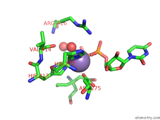
Mono view
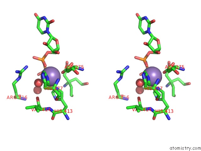
Stereo pair view
Mono view
Stereo pair view
A full contact list of Manganese with other atoms in the Mn binding
site number 1 of Crystal Structure of the Tggalnac-T3 in Complex with Udp, Manganese and the Peptide 3 within 5.0Å range:
|
Reference:
M.De Las Rivas,
E.J.P.Daniel,
Y.Narimatsu,
I.Companon,
K.Kato,
P.Hermosilla,
A.Thureau,
L.Ceballos-Laita,
H.Coelho,
P.Bernado,
F.Marcelo,
L.Hansen,
A.Lostao,
F.Corzana,
H.Clausen,
T.A.Gerken,
R.Hurtado-Guerrero.
Molecular Basis For Fibroblast Growth Factor 23 O-Glycosylation By Galnac-T3. Nat.Chem.Biol. 2019.
ISSN: ESSN 1552-4469
Page generated: Sun Oct 6 07:05:11 2024
ISSN: ESSN 1552-4469
Last articles
F in 4IBIF in 4IAH
F in 4IAE
F in 4I9H
F in 4I9N
F in 4I9O
F in 4IA9
F in 4I89
F in 4I7S
F in 4I87