Atomistry » Manganese » PDB 6oe2-6q9f » 6q92
Atomistry »
  Manganese »
    PDB 6oe2-6q9f »
      6q92 »

Manganese in PDB 6q92: Crystal Structure of Human Arginase-1 at pH 7.0 in Complex with Abh

Enzymatic activity of Crystal Structure of Human Arginase-1 at pH 7.0 in Complex with Abh

All present enzymatic activity of Crystal Structure of Human Arginase-1 at pH 7.0 in Complex with Abh:
3.5.3.1;

Protein crystallography data

The structure of Crystal Structure of Human Arginase-1 at pH 7.0 in Complex with Abh, PDB code: 6q92 was solved by Y.Grobben, J.C.M.Uitdehaag, G.J.R.Zaman, with X-Ray Crystallography technique. A brief refinement statistics is given in the table below:

Resolution Low / High (Å) 39.14 / 1.50
Space group P 3
Cell size a, b, c (Å), α, β, γ (°) 90.380, 90.380, 69.254, 90.00, 90.00, 120.00
R / Rfree (%) 13.5 / 14.7

Other elements in 6q92:

The structure of Crystal Structure of Human Arginase-1 at pH 7.0 in Complex with Abh also contains other interesting chemical elements:

Sodium (Na) 2 atoms

Manganese Binding Sites:

The binding sites of Manganese atom in the Crystal Structure of Human Arginase-1 at pH 7.0 in Complex with Abh (pdb code 6q92). This binding sites where shown within 5.0 Angstroms radius around Manganese atom.
In total 4 binding sites of Manganese where determined in the Crystal Structure of Human Arginase-1 at pH 7.0 in Complex with Abh, PDB code: 6q92:
Jump to Manganese binding site number: 1; 2; 3; 4;

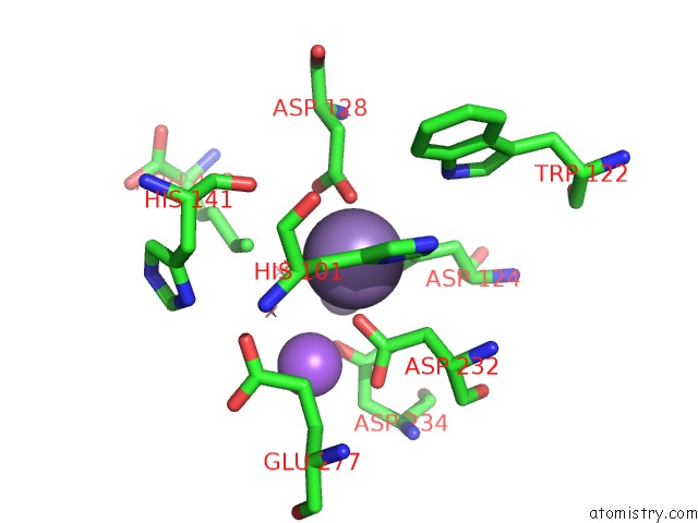
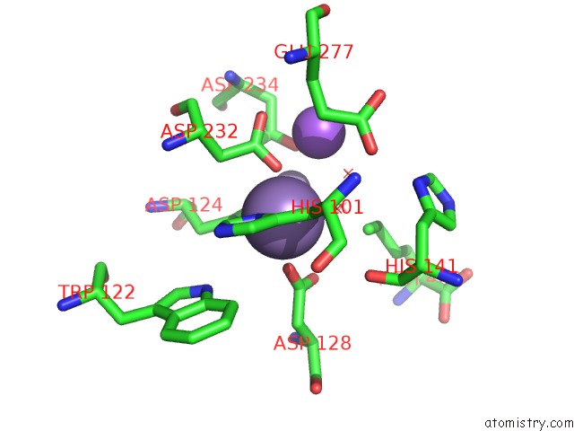
Manganese binding site 1 out of 4 in 6q92

Go back to Manganese Binding Sites List in 6q92
Manganese binding site 1 out of 4 in the Crystal Structure of Human Arginase-1 at pH 7.0 in Complex with Abh


Mono view


Stereo pair view

A full contact list of Manganese with other atoms in the Mn binding site number 1 of Crystal Structure of Human Arginase-1 at pH 7.0 in Complex with Abh within 5.0Å range:
probe atom residue distance (Å) B Occ
A:Mn401

b:7.8
occ:1.00
OD2 A:ASP128 2.1 8.7 1.0
OD2 A:ASP232 2.2 7.2 1.0
O2 A:ABH403 2.2 11.0 1.0
OD2 A:ASP124 2.2 7.3 1.0
ND1 A:HIS101 2.2 7.6 1.0
O1 A:ABH403 2.3 9.7 1.0
B A:ABH403 2.8 9.9 1.0
CG A:ASP128 3.1 8.1 1.0
CG A:ASP124 3.2 6.5 1.0
CG A:HIS101 3.2 8.0 1.0
CG A:ASP232 3.2 7.3 1.0
CE1 A:HIS101 3.3 8.1 1.0
OD1 A:ASP128 3.3 8.6 1.0
CB A:HIS101 3.4 8.1 1.0
MN A:MN402 3.4 6.2 1.0
OD1 A:ASP124 3.5 6.7 1.0
CB A:ASP232 3.6 7.3 1.0
O3 A:ABH403 3.7 9.4 1.0
CE A:ABH403 4.1 10.2 1.0
NA A:NA404 4.2 12.2 1.0
CD2 A:HIS101 4.3 8.0 1.0
NE2 A:HIS101 4.3 8.0 1.0
OD1 A:ASP232 4.4 7.4 1.0
NE1 A:TRP122 4.4 6.8 1.0
O A:HIS141 4.4 8.3 1.0
CB A:ASP128 4.5 7.8 1.0
CB A:ASP124 4.5 6.3 1.0
OD2 A:ASP234 4.6 8.7 1.0
CZ2 A:TRP122 4.6 6.8 1.0
CE2 A:TRP122 4.9 6.9 1.0
CG A:GLU277 4.9 9.9 1.0
CA A:HIS101 4.9 7.9 1.0
CA A:ASP232 5.0 7.4 1.0
OE2 A:GLU277 5.0 11.0 1.0

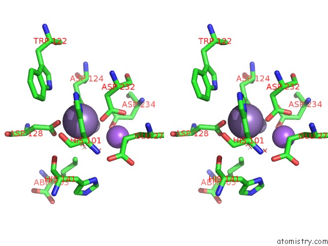
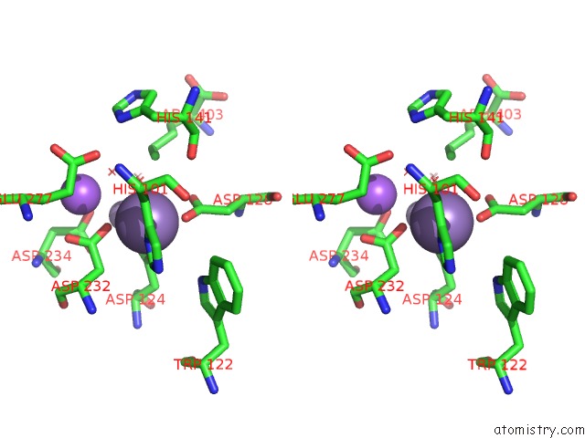
Manganese binding site 2 out of 4 in 6q92

Go back to Manganese Binding Sites List in 6q92
Manganese binding site 2 out of 4 in the Crystal Structure of Human Arginase-1 at pH 7.0 in Complex with Abh


Mono view


Stereo pair view

A full contact list of Manganese with other atoms in the Mn binding site number 2 of Crystal Structure of Human Arginase-1 at pH 7.0 in Complex with Abh within 5.0Å range:
probe atom residue distance (Å) B Occ
A:Mn402

b:6.2
occ:1.00
O1 A:ABH403 1.9 9.7 1.0
OD1 A:ASP124 2.0 6.7 1.0
ND1 A:HIS126 2.0 8.2 1.0
OD2 A:ASP234 2.0 8.7 1.0
CG A:ASP234 2.8 7.7 1.0
OD1 A:ASP234 2.8 7.4 1.0
CE1 A:HIS126 2.9 7.6 1.0
OD2 A:ASP232 3.0 7.2 1.0
CG A:ASP124 3.0 6.5 1.0
CG A:HIS126 3.0 7.6 1.0
B A:ABH403 3.1 9.9 1.0
O3 A:ABH403 3.1 9.4 1.0
NA A:NA404 3.4 12.2 1.0
CB A:HIS126 3.4 7.4 1.0
MN A:MN401 3.4 7.8 1.0
OD2 A:ASP124 3.4 7.3 1.0
CG A:ASP232 3.7 7.3 1.0
N A:HIS126 3.8 6.8 1.0
O2 A:ABH403 4.0 11.0 1.0
OG1 A:THR246 4.0 11.5 1.0
NE2 A:HIS126 4.1 8.0 1.0
CE A:ABH403 4.1 10.2 1.0
OD1 A:ASP128 4.1 8.6 1.0
N A:ALA125 4.1 6.3 1.0
CD2 A:HIS126 4.1 7.8 1.0
OD1 A:ASP232 4.2 7.4 1.0
CD A:ABH403 4.2 10.3 1.0
CA A:HIS126 4.2 7.1 1.0
CB A:ASP234 4.2 7.6 1.0
CB A:ASP124 4.4 6.3 1.0
CB A:ASP232 4.5 7.3 1.0
CB A:ALA125 4.5 6.6 1.0
C A:ALA125 4.6 6.5 1.0
CA A:ALA125 4.6 6.5 1.0
OD2 A:ASP128 4.7 8.7 1.0
CA A:ASP124 4.8 6.4 1.0
CG A:ASP128 4.8 8.1 1.0
O A:HIS126 4.8 7.5 1.0
C A:ASP124 4.9 6.3 1.0
C A:HIS126 5.0 7.0 1.0
O A:THR246 5.0 10.2 1.0

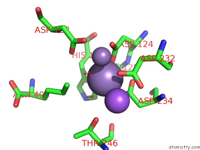
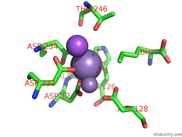
Manganese binding site 3 out of 4 in 6q92

Go back to Manganese Binding Sites List in 6q92
Manganese binding site 3 out of 4 in the Crystal Structure of Human Arginase-1 at pH 7.0 in Complex with Abh


Mono view


Stereo pair view

A full contact list of Manganese with other atoms in the Mn binding site number 3 of Crystal Structure of Human Arginase-1 at pH 7.0 in Complex with Abh within 5.0Å range:
probe atom residue distance (Å) B Occ
B:Mn401

b:9.6
occ:1.00
OD2 B:ASP124 2.1 7.4 1.0
OD2 B:ASP128 2.1 9.2 1.0
OD2 B:ASP232 2.2 8.7 1.0
ND1 B:HIS101 2.2 10.3 1.0
O1 B:ABH403 2.3 13.3 1.0
O2 B:ABH403 2.3 13.4 1.0
B B:ABH403 2.8 12.3 1.0
CG B:ASP124 3.1 7.3 1.0
CG B:ASP128 3.1 8.9 1.0
CE1 B:HIS101 3.2 10.3 1.0
CG B:HIS101 3.2 10.6 1.0
CG B:ASP232 3.3 9.0 1.0
MN B:MN402 3.4 7.8 1.0
OD1 B:ASP128 3.4 9.7 1.0
OD1 B:ASP124 3.5 7.7 1.0
CB B:HIS101 3.5 11.0 1.0
CB B:ASP232 3.6 9.2 1.0
O3 B:ABH403 3.7 11.8 1.0
CE B:ABH403 4.0 12.9 1.0
NA B:NA404 4.1 14.1 1.0
NE2 B:HIS101 4.3 10.1 1.0
CD2 B:HIS101 4.3 10.6 1.0
NE1 B:TRP122 4.3 9.3 1.0
OD1 B:ASP232 4.4 8.8 1.0
O B:HIS141 4.4 11.6 1.0
CB B:ASP124 4.5 7.7 1.0
CB B:ASP128 4.5 8.9 1.0
CZ2 B:TRP122 4.6 9.5 1.0
OD2 B:ASP234 4.6 10.4 1.0
CE2 B:TRP122 4.8 9.2 1.0
CG B:GLU277 4.9 13.0 1.0
CA B:ASP232 5.0 9.4 1.0
OE2 B:GLU277 5.0 14.1 1.0

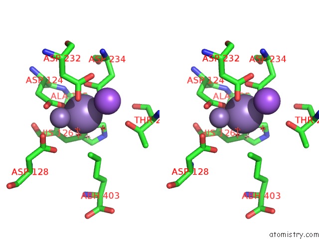
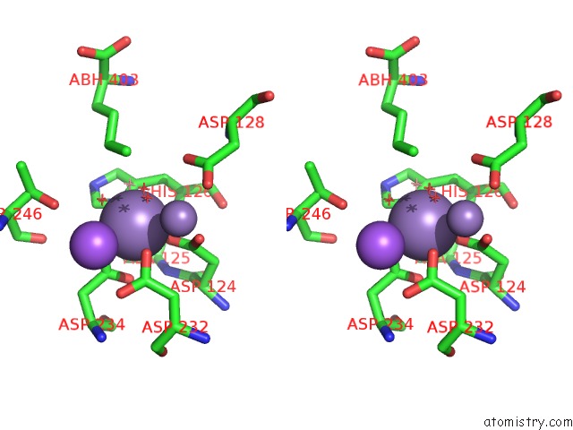
Manganese binding site 4 out of 4 in 6q92

Go back to Manganese Binding Sites List in 6q92
Manganese binding site 4 out of 4 in the Crystal Structure of Human Arginase-1 at pH 7.0 in Complex with Abh


Mono view


Stereo pair view

A full contact list of Manganese with other atoms in the Mn binding site number 4 of Crystal Structure of Human Arginase-1 at pH 7.0 in Complex with Abh within 5.0Å range:
probe atom residue distance (Å) B Occ
B:Mn402

b:7.8
occ:1.00
O1 B:ABH403 1.9 13.3 1.0
OD2 B:ASP234 2.0 10.4 1.0
OD1 B:ASP124 2.0 7.7 1.0
ND1 B:HIS126 2.0 9.2 1.0
CG B:ASP234 2.7 9.2 1.0
OD1 B:ASP234 2.8 10.1 1.0
CE1 B:HIS126 2.9 9.0 1.0
CG B:ASP124 3.0 7.3 1.0
OD2 B:ASP232 3.0 8.7 1.0
B B:ABH403 3.0 12.3 1.0
CG B:HIS126 3.1 9.1 1.0
O3 B:ABH403 3.1 11.8 1.0
NA B:NA404 3.3 14.1 1.0
OD2 B:ASP124 3.4 7.4 1.0
CB B:HIS126 3.4 8.8 1.0
MN B:MN401 3.4 9.6 1.0
CG B:ASP232 3.7 9.0 1.0
N B:HIS126 3.9 8.1 1.0
O2 B:ABH403 4.0 13.4 1.0
CE B:ABH403 4.0 12.9 1.0
OG1 B:THR246 4.1 13.3 1.0
NE2 B:HIS126 4.1 9.5 1.0
CD B:ABH403 4.1 13.2 1.0
N B:ALA125 4.1 7.6 1.0
CD2 B:HIS126 4.2 9.8 1.0
OD1 B:ASP232 4.2 8.8 1.0
OD1 B:ASP128 4.2 9.7 1.0
CB B:ASP234 4.2 9.2 1.0
CA B:HIS126 4.3 8.6 1.0
CB B:ASP124 4.4 7.7 1.0
CB B:ASP232 4.5 9.2 1.0
CB B:ALA125 4.6 7.5 1.0
OD2 B:ASP128 4.6 9.2 1.0
C B:ALA125 4.7 7.6 1.0
CA B:ALA125 4.7 7.6 1.0
CA B:ASP124 4.8 7.7 1.0
CG B:ASP128 4.8 8.9 1.0
C B:ASP124 4.9 7.6 1.0
O B:THR246 4.9 12.8 1.0
O B:HIS126 4.9 9.0 1.0

Reference:

Y.Grobben, J.C.M.Uitdehaag, G.J.R.Zaman. Structural Insights Into Human Arginase-1 pH Dependence and Its Inhibition By the Small Molecule Inhibitor Cb-1158 J.Struct.Biol. 2019.
ISSN: ESSN 1095-8657
DOI: 10.1016/J.YJSBX.2019.100014
Page generated: Sun Oct 6 05:54:44 2024

Last articles

Zn in 9J0N
Zn in 9J0O
Zn in 9J0P
Zn in 9FJX
Zn in 9EKB
Zn in 9C0F
Zn in 9CAH
Zn in 9CH0
Zn in 9CH3
Zn in 9CH1
© Copyright 2008-2020 by atomistry.com
Home   |    Site Map   |    Copyright   |    Contact us   |    Privacy