Manganese »
PDB 6oe2-6q9f »
6ozq »
Manganese in PDB 6ozq: Crystal Structure of Mus Musculus (Mm) Endonuclease V (K155M) in Complex with A 23MER Rna Oligo Containing An Inosine After A 100 Min Soak in 10 Mm MN2+ and K+
Protein crystallography data
The structure of Crystal Structure of Mus Musculus (Mm) Endonuclease V (K155M) in Complex with A 23MER Rna Oligo Containing An Inosine After A 100 Min Soak in 10 Mm MN2+ and K+, PDB code: 6ozq
was solved by
N.L.Samara,
W.Yang,
with X-Ray Crystallography technique. A brief refinement statistics is given in the table below:
| Resolution Low / High (Å) | 42.72 / 2.15 |
| Space group | P 21 21 21 |
| Cell size a, b, c (Å), α, β, γ (°) | 71.205, 73.807, 154.659, 90.00, 90.00, 90.00 |
| R / Rfree (%) | 17.4 / 20.9 |
Manganese Binding Sites:
The binding sites of Manganese atom in the Crystal Structure of Mus Musculus (Mm) Endonuclease V (K155M) in Complex with A 23MER Rna Oligo Containing An Inosine After A 100 Min Soak in 10 Mm MN2+ and K+
(pdb code 6ozq). This binding sites where shown within
5.0 Angstroms radius around Manganese atom.
In total 7 binding sites of Manganese where determined in the Crystal Structure of Mus Musculus (Mm) Endonuclease V (K155M) in Complex with A 23MER Rna Oligo Containing An Inosine After A 100 Min Soak in 10 Mm MN2+ and K+, PDB code: 6ozq:
Jump to Manganese binding site number: 1; 2; 3; 4; 5; 6; 7;
In total 7 binding sites of Manganese where determined in the Crystal Structure of Mus Musculus (Mm) Endonuclease V (K155M) in Complex with A 23MER Rna Oligo Containing An Inosine After A 100 Min Soak in 10 Mm MN2+ and K+, PDB code: 6ozq:
Jump to Manganese binding site number: 1; 2; 3; 4; 5; 6; 7;
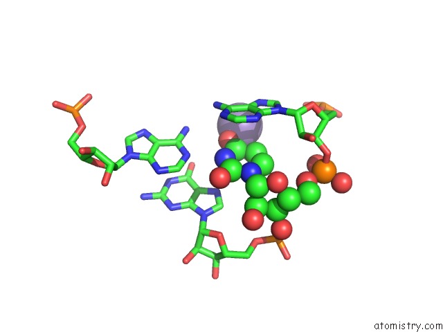
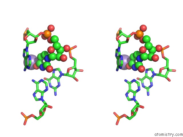
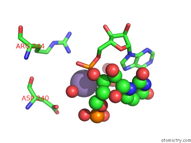
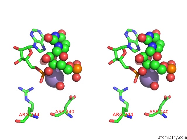
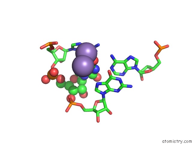
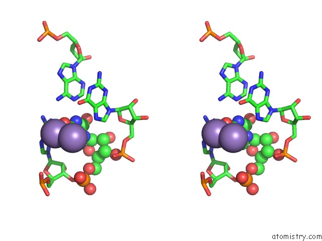
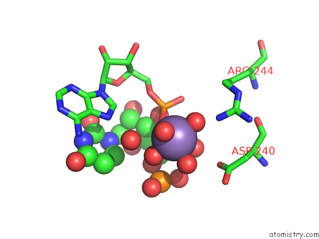
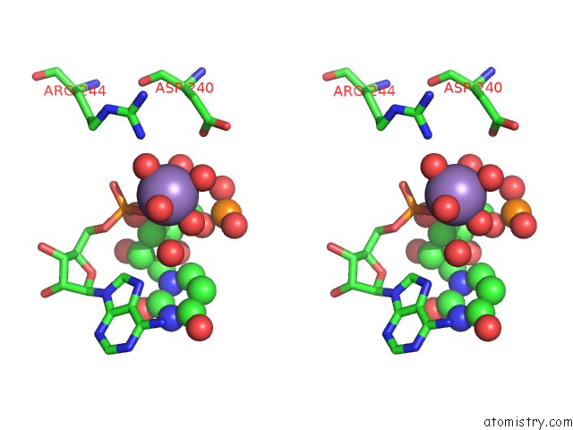
Manganese binding site 1 out of 7 in 6ozq
Go back to
Manganese binding site 1 out
of 7 in the Crystal Structure of Mus Musculus (Mm) Endonuclease V (K155M) in Complex with A 23MER Rna Oligo Containing An Inosine After A 100 Min Soak in 10 Mm MN2+ and K+
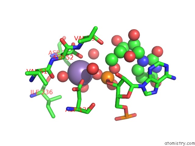
Mono view
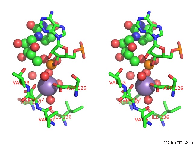
Stereo pair view
Mono view
Stereo pair view
A full contact list of Manganese with other atoms in the Mn binding
site number 1 of Crystal Structure of Mus Musculus (Mm) Endonuclease V (K155M) in Complex with A 23MER Rna Oligo Containing An Inosine After A 100 Min Soak in 10 Mm MN2+ and K+ within 5.0Å range:
|
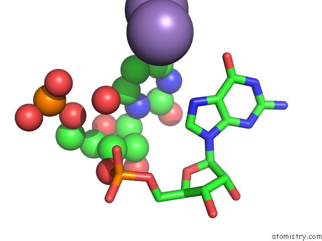
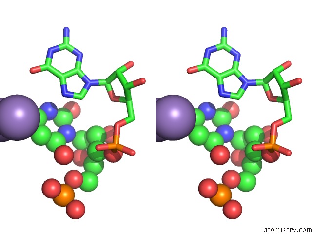
Manganese binding site 2 out of 7 in 6ozq
Go back to
Manganese binding site 2 out
of 7 in the Crystal Structure of Mus Musculus (Mm) Endonuclease V (K155M) in Complex with A 23MER Rna Oligo Containing An Inosine After A 100 Min Soak in 10 Mm MN2+ and K+
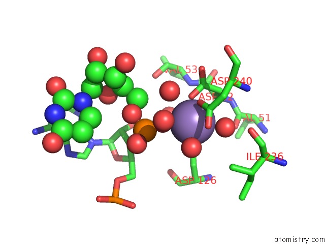
Mono view
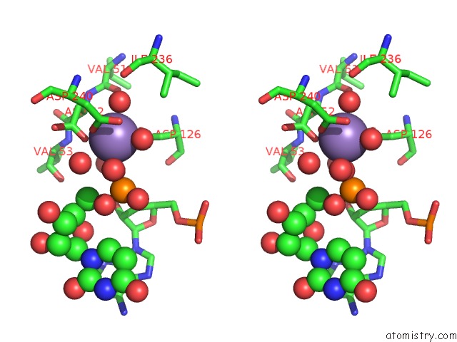
Stereo pair view
Mono view
Stereo pair view
A full contact list of Manganese with other atoms in the Mn binding
site number 2 of Crystal Structure of Mus Musculus (Mm) Endonuclease V (K155M) in Complex with A 23MER Rna Oligo Containing An Inosine After A 100 Min Soak in 10 Mm MN2+ and K+ within 5.0Å range:
|
Manganese binding site 3 out of 7 in 6ozq
Go back to
Manganese binding site 3 out
of 7 in the Crystal Structure of Mus Musculus (Mm) Endonuclease V (K155M) in Complex with A 23MER Rna Oligo Containing An Inosine After A 100 Min Soak in 10 Mm MN2+ and K+
Mono view
Stereo pair view
Mono view
Stereo pair view
A full contact list of Manganese with other atoms in the Mn binding
site number 3 of Crystal Structure of Mus Musculus (Mm) Endonuclease V (K155M) in Complex with A 23MER Rna Oligo Containing An Inosine After A 100 Min Soak in 10 Mm MN2+ and K+ within 5.0Å range:
|
Manganese binding site 4 out of 7 in 6ozq
Go back to
Manganese binding site 4 out
of 7 in the Crystal Structure of Mus Musculus (Mm) Endonuclease V (K155M) in Complex with A 23MER Rna Oligo Containing An Inosine After A 100 Min Soak in 10 Mm MN2+ and K+
Mono view
Stereo pair view
Mono view
Stereo pair view
A full contact list of Manganese with other atoms in the Mn binding
site number 4 of Crystal Structure of Mus Musculus (Mm) Endonuclease V (K155M) in Complex with A 23MER Rna Oligo Containing An Inosine After A 100 Min Soak in 10 Mm MN2+ and K+ within 5.0Å range:
|
Manganese binding site 5 out of 7 in 6ozq
Go back to
Manganese binding site 5 out
of 7 in the Crystal Structure of Mus Musculus (Mm) Endonuclease V (K155M) in Complex with A 23MER Rna Oligo Containing An Inosine After A 100 Min Soak in 10 Mm MN2+ and K+
Mono view
Stereo pair view
Mono view
Stereo pair view
A full contact list of Manganese with other atoms in the Mn binding
site number 5 of Crystal Structure of Mus Musculus (Mm) Endonuclease V (K155M) in Complex with A 23MER Rna Oligo Containing An Inosine After A 100 Min Soak in 10 Mm MN2+ and K+ within 5.0Å range:
|
Manganese binding site 6 out of 7 in 6ozq
Go back to
Manganese binding site 6 out
of 7 in the Crystal Structure of Mus Musculus (Mm) Endonuclease V (K155M) in Complex with A 23MER Rna Oligo Containing An Inosine After A 100 Min Soak in 10 Mm MN2+ and K+
Mono view
Stereo pair view
Mono view
Stereo pair view
A full contact list of Manganese with other atoms in the Mn binding
site number 6 of Crystal Structure of Mus Musculus (Mm) Endonuclease V (K155M) in Complex with A 23MER Rna Oligo Containing An Inosine After A 100 Min Soak in 10 Mm MN2+ and K+ within 5.0Å range:
|
Manganese binding site 7 out of 7 in 6ozq
Go back to
Manganese binding site 7 out
of 7 in the Crystal Structure of Mus Musculus (Mm) Endonuclease V (K155M) in Complex with A 23MER Rna Oligo Containing An Inosine After A 100 Min Soak in 10 Mm MN2+ and K+
Mono view
Stereo pair view
Mono view
Stereo pair view
A full contact list of Manganese with other atoms in the Mn binding
site number 7 of Crystal Structure of Mus Musculus (Mm) Endonuclease V (K155M) in Complex with A 23MER Rna Oligo Containing An Inosine After A 100 Min Soak in 10 Mm MN2+ and K+ within 5.0Å range:
|
Reference:
J.Wu,
N.L.Samara,
I.Kuraoka,
W.Yang.
Evolution of Inosine-Specific Endonuclease V From Bacterial Dnase to Eukaryotic Rnase. Mol.Cell V. 76 44 2019.
ISSN: ISSN 1097-2765
PubMed: 31444105
DOI: 10.1016/J.MOLCEL.2019.06.046
Page generated: Sun Oct 6 05:48:06 2024
ISSN: ISSN 1097-2765
PubMed: 31444105
DOI: 10.1016/J.MOLCEL.2019.06.046
Last articles
Zn in 9MJ5Zn in 9HNW
Zn in 9G0L
Zn in 9FNE
Zn in 9DZN
Zn in 9E0I
Zn in 9D32
Zn in 9DAK
Zn in 8ZXC
Zn in 8ZUF