Manganese »
PDB 6oe2-6q9f »
6omr »
Manganese in PDB 6omr: Crystal Structure of PTMU3 Complexed with Ptn Substrate
Protein crystallography data
The structure of Crystal Structure of PTMU3 Complexed with Ptn Substrate, PDB code: 6omr
was solved by
Y.C.Liu,
L.B.Dong,
B.Shen,
with X-Ray Crystallography technique. A brief refinement statistics is given in the table below:
| Resolution Low / High (Å) | 29.83 / 2.45 |
| Space group | I 2 2 2 |
| Cell size a, b, c (Å), α, β, γ (°) | 113.620, 122.420, 140.063, 90.00, 90.00, 90.00 |
| R / Rfree (%) | 18.2 / 25.1 |
Manganese Binding Sites:
The binding sites of Manganese atom in the Crystal Structure of PTMU3 Complexed with Ptn Substrate
(pdb code 6omr). This binding sites where shown within
5.0 Angstroms radius around Manganese atom.
In total 5 binding sites of Manganese where determined in the Crystal Structure of PTMU3 Complexed with Ptn Substrate, PDB code: 6omr:
Jump to Manganese binding site number: 1; 2; 3; 4; 5;
In total 5 binding sites of Manganese where determined in the Crystal Structure of PTMU3 Complexed with Ptn Substrate, PDB code: 6omr:
Jump to Manganese binding site number: 1; 2; 3; 4; 5;
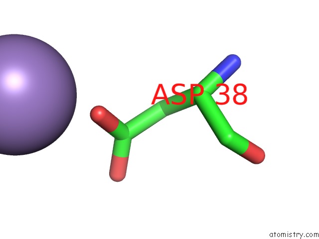
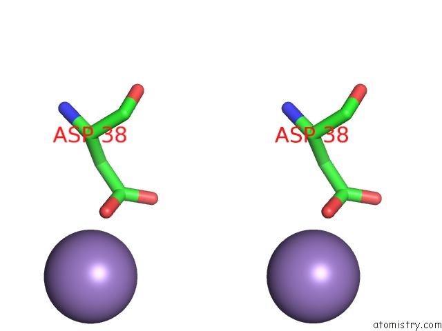
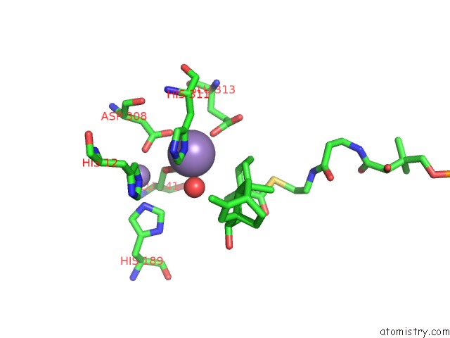
Manganese binding site 1 out of 5 in 6omr
Go back to
Manganese binding site 1 out
of 5 in the Crystal Structure of PTMU3 Complexed with Ptn Substrate
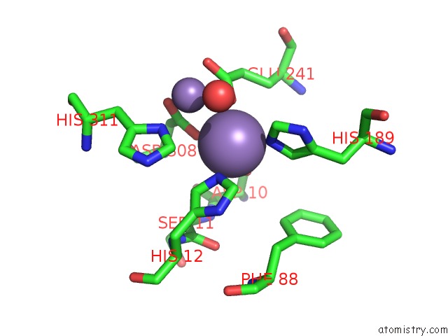
Mono view
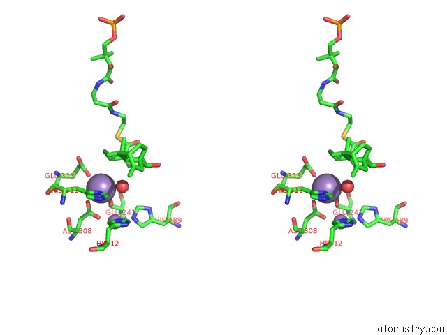
Stereo pair view
Mono view
Stereo pair view
A full contact list of Manganese with other atoms in the Mn binding
site number 1 of Crystal Structure of PTMU3 Complexed with Ptn Substrate within 5.0Å range:
|
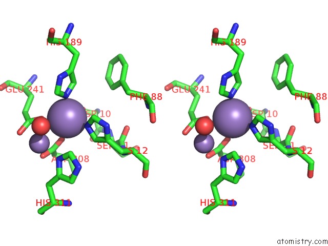
Manganese binding site 2 out of 5 in 6omr
Go back to
Manganese binding site 2 out
of 5 in the Crystal Structure of PTMU3 Complexed with Ptn Substrate
Mono view
Stereo pair view
Mono view
Stereo pair view
A full contact list of Manganese with other atoms in the Mn binding
site number 2 of Crystal Structure of PTMU3 Complexed with Ptn Substrate within 5.0Å range:
|
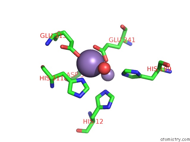
Manganese binding site 3 out of 5 in 6omr
Go back to
Manganese binding site 3 out
of 5 in the Crystal Structure of PTMU3 Complexed with Ptn Substrate
Mono view
Stereo pair view
Mono view
Stereo pair view
A full contact list of Manganese with other atoms in the Mn binding
site number 3 of Crystal Structure of PTMU3 Complexed with Ptn Substrate within 5.0Å range:
|
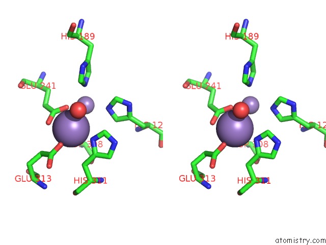
Manganese binding site 4 out of 5 in 6omr
Go back to
Manganese binding site 4 out
of 5 in the Crystal Structure of PTMU3 Complexed with Ptn Substrate
Mono view
Stereo pair view
Mono view
Stereo pair view
A full contact list of Manganese with other atoms in the Mn binding
site number 4 of Crystal Structure of PTMU3 Complexed with Ptn Substrate within 5.0Å range:
|
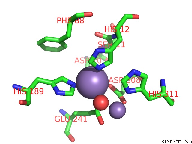
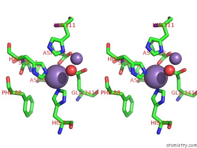
Manganese binding site 5 out of 5 in 6omr
Go back to
Manganese binding site 5 out
of 5 in the Crystal Structure of PTMU3 Complexed with Ptn Substrate
Mono view
Stereo pair view
Mono view
Stereo pair view
A full contact list of Manganese with other atoms in the Mn binding
site number 5 of Crystal Structure of PTMU3 Complexed with Ptn Substrate within 5.0Å range:
|
Reference:
L.B.Dong,
Y.C.Liu,
A.J.Cepeda,
E.Kalkreuter,
M.R.Deng,
J.D.Rudolf,
C.Chang,
A.Joachimiak,
G.N.Phillips Jr.,
B.Shen.
Characterization and Crystal Structure of A Nonheme Diiron Monooxygenase Involved in Platensimycin and Platencin Biosynthesis. J.Am.Chem.Soc. V. 141 12406 2019.
ISSN: ESSN 1520-5126
PubMed: 31291107
DOI: 10.1021/JACS.9B06183
Page generated: Sun Oct 6 05:43:51 2024
ISSN: ESSN 1520-5126
PubMed: 31291107
DOI: 10.1021/JACS.9B06183
Last articles
Zn in 9J0NZn in 9J0O
Zn in 9J0P
Zn in 9FJX
Zn in 9EKB
Zn in 9C0F
Zn in 9CAH
Zn in 9CH0
Zn in 9CH3
Zn in 9CH1