Manganese »
PDB 6oe2-6q9f »
6oiy »
Manganese in PDB 6oiy: Structure of Escherichia Coli Bound to Dgtp
Enzymatic activity of Structure of Escherichia Coli Bound to Dgtp
All present enzymatic activity of Structure of Escherichia Coli Bound to Dgtp:
3.1.5.1;
3.1.5.1;
Protein crystallography data
The structure of Structure of Escherichia Coli Bound to Dgtp, PDB code: 6oiy
was solved by
C.O.Barnes,
Y.Wu,
G.Calero,
with X-Ray Crystallography technique. A brief refinement statistics is given in the table below:
| Resolution Low / High (Å) | 46.87 / 3.29 |
| Space group | P 43 21 2 |
| Cell size a, b, c (Å), α, β, γ (°) | 192.176, 192.176, 299.596, 90.00, 90.00, 90.00 |
| R / Rfree (%) | 20.5 / 24.1 |
Manganese Binding Sites:
The binding sites of Manganese atom in the Structure of Escherichia Coli Bound to Dgtp
(pdb code 6oiy). This binding sites where shown within
5.0 Angstroms radius around Manganese atom.
In total 6 binding sites of Manganese where determined in the Structure of Escherichia Coli Bound to Dgtp, PDB code: 6oiy:
Jump to Manganese binding site number: 1; 2; 3; 4; 5; 6;
In total 6 binding sites of Manganese where determined in the Structure of Escherichia Coli Bound to Dgtp, PDB code: 6oiy:
Jump to Manganese binding site number: 1; 2; 3; 4; 5; 6;
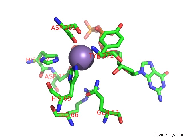
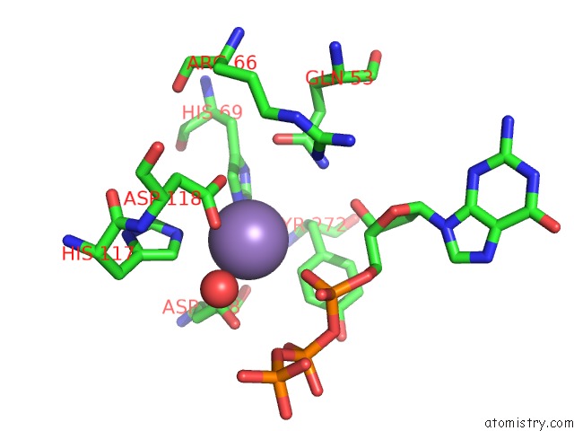
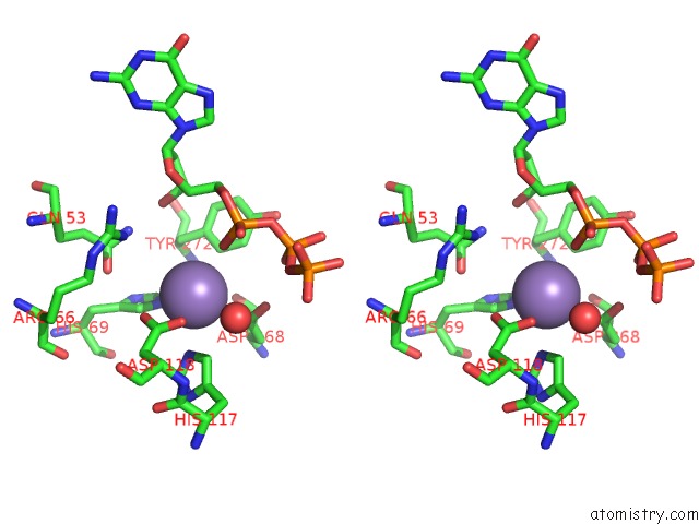
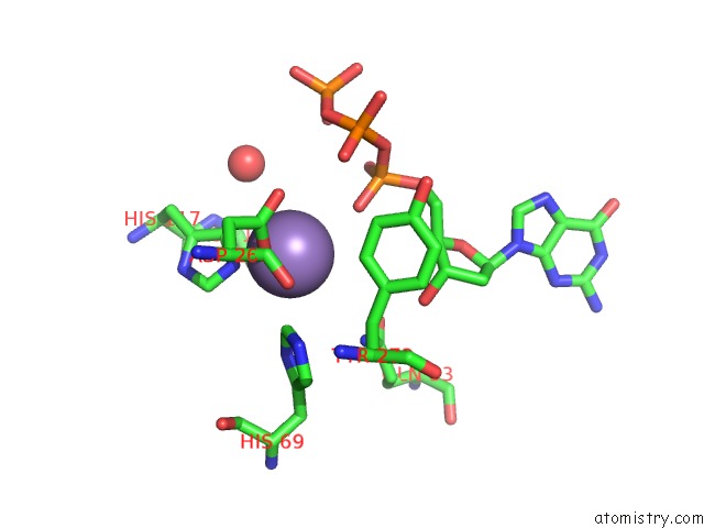
Manganese binding site 1 out of 6 in 6oiy
Go back to
Manganese binding site 1 out
of 6 in the Structure of Escherichia Coli Bound to Dgtp
Mono view
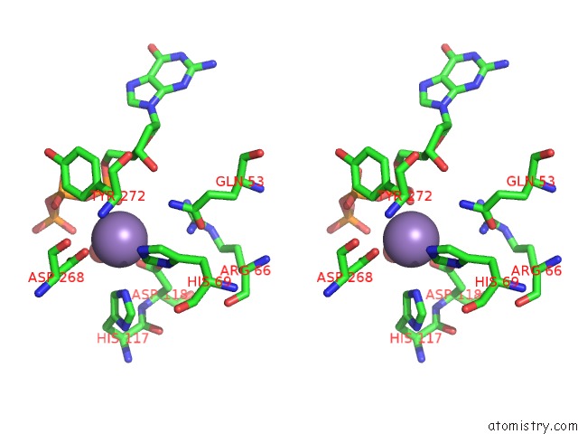
Stereo pair view
Mono view
Stereo pair view
A full contact list of Manganese with other atoms in the Mn binding
site number 1 of Structure of Escherichia Coli Bound to Dgtp within 5.0Å range:
|
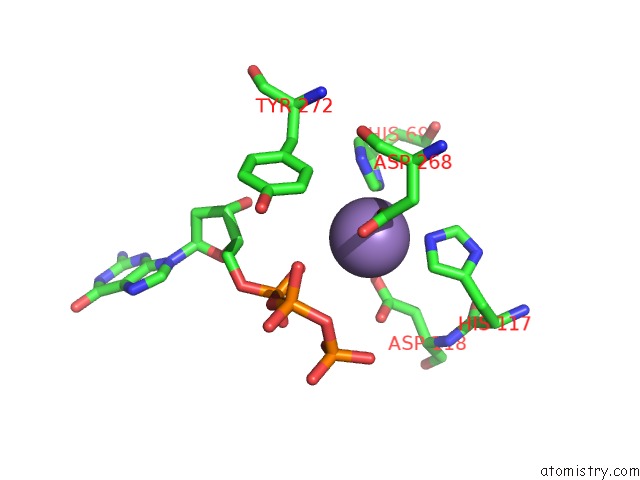
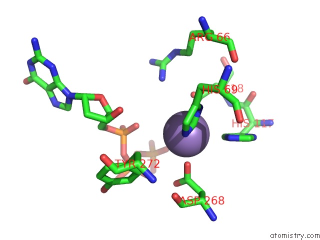
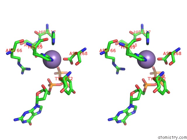
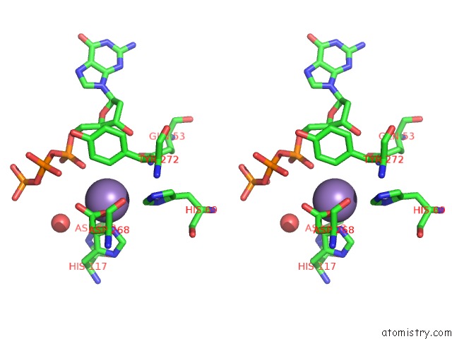
Manganese binding site 2 out of 6 in 6oiy
Go back to
Manganese binding site 2 out
of 6 in the Structure of Escherichia Coli Bound to Dgtp
Mono view
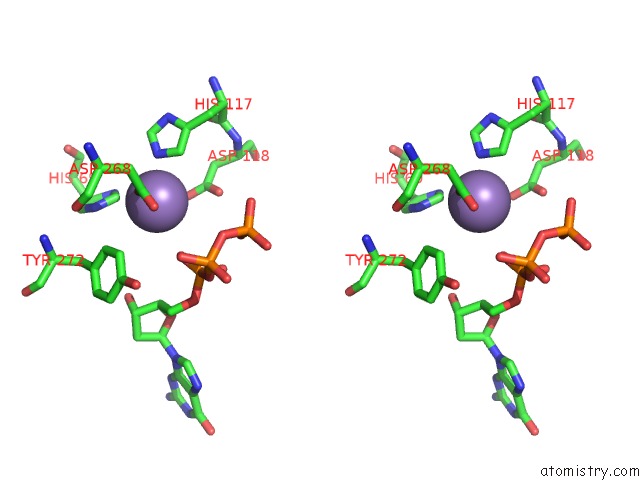
Stereo pair view
Mono view
Stereo pair view
A full contact list of Manganese with other atoms in the Mn binding
site number 2 of Structure of Escherichia Coli Bound to Dgtp within 5.0Å range:
|
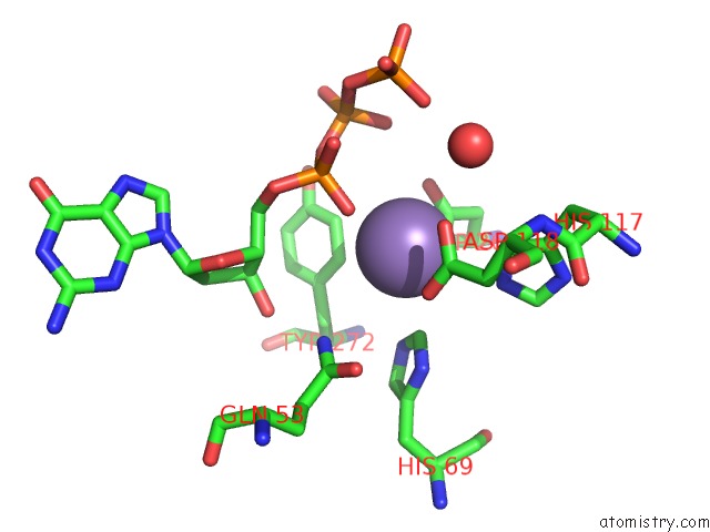
Manganese binding site 3 out of 6 in 6oiy
Go back to
Manganese binding site 3 out
of 6 in the Structure of Escherichia Coli Bound to Dgtp
Mono view
Stereo pair view
Mono view
Stereo pair view
A full contact list of Manganese with other atoms in the Mn binding
site number 3 of Structure of Escherichia Coli Bound to Dgtp within 5.0Å range:
|
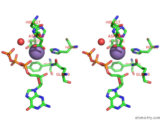
Manganese binding site 4 out of 6 in 6oiy
Go back to
Manganese binding site 4 out
of 6 in the Structure of Escherichia Coli Bound to Dgtp
Mono view
Stereo pair view
Mono view
Stereo pair view
A full contact list of Manganese with other atoms in the Mn binding
site number 4 of Structure of Escherichia Coli Bound to Dgtp within 5.0Å range:
|
Manganese binding site 5 out of 6 in 6oiy
Go back to
Manganese binding site 5 out
of 6 in the Structure of Escherichia Coli Bound to Dgtp
Mono view
Stereo pair view
Mono view
Stereo pair view
A full contact list of Manganese with other atoms in the Mn binding
site number 5 of Structure of Escherichia Coli Bound to Dgtp within 5.0Å range:
|
Manganese binding site 6 out of 6 in 6oiy
Go back to
Manganese binding site 6 out
of 6 in the Structure of Escherichia Coli Bound to Dgtp
Mono view
Stereo pair view
Mono view
Stereo pair view
A full contact list of Manganese with other atoms in the Mn binding
site number 6 of Structure of Escherichia Coli Bound to Dgtp within 5.0Å range:
|
Reference:
C.O.Barnes,
Y.Wu,
J.Song,
G.Lin,
E.L.Baxter,
A.S.Brewster,
V.Nagarajan,
A.Holmes,
S.M.Soltis,
N.K.Sauter,
J.Ahn,
A.E.Cohen,
G.Calero.
The Crystal Structure of Dgtpase Reveals the Molecular Basis of Dgtp Selectivity. Proc.Natl.Acad.Sci.Usa V. 116 9333 2019.
ISSN: ESSN 1091-6490
PubMed: 31019074
DOI: 10.1073/PNAS.1814999116
Page generated: Sun Oct 6 05:43:05 2024
ISSN: ESSN 1091-6490
PubMed: 31019074
DOI: 10.1073/PNAS.1814999116
Last articles
Fe in 2YXOFe in 2YRS
Fe in 2YXC
Fe in 2YNM
Fe in 2YVJ
Fe in 2YP1
Fe in 2YU2
Fe in 2YU1
Fe in 2YQB
Fe in 2YOO