Manganese »
PDB 6juq-6luh »
6lrd »
Manganese in PDB 6lrd: Structure of Recj Complexed with A 5'-P-Dspacer-Modified Ssdna
Protein crystallography data
The structure of Structure of Recj Complexed with A 5'-P-Dspacer-Modified Ssdna, PDB code: 6lrd
was solved by
K.Cheng,
Y.Hua,
with X-Ray Crystallography technique. A brief refinement statistics is given in the table below:
| Resolution Low / High (Å) | 29.35 / 1.90 |
| Space group | P 32 2 1 |
| Cell size a, b, c (Å), α, β, γ (°) | 106.140, 106.140, 164.560, 90.00, 90.00, 120.00 |
| R / Rfree (%) | 20.8 / 23.4 |
Manganese Binding Sites:
The binding sites of Manganese atom in the Structure of Recj Complexed with A 5'-P-Dspacer-Modified Ssdna
(pdb code 6lrd). This binding sites where shown within
5.0 Angstroms radius around Manganese atom.
In total 2 binding sites of Manganese where determined in the Structure of Recj Complexed with A 5'-P-Dspacer-Modified Ssdna, PDB code: 6lrd:
Jump to Manganese binding site number: 1; 2;
In total 2 binding sites of Manganese where determined in the Structure of Recj Complexed with A 5'-P-Dspacer-Modified Ssdna, PDB code: 6lrd:
Jump to Manganese binding site number: 1; 2;
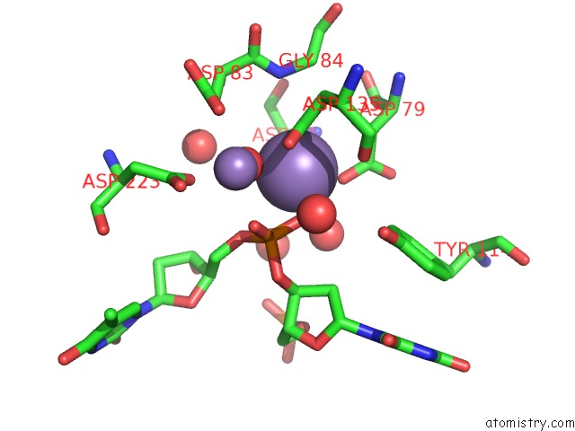
Manganese binding site 1 out of 2 in 6lrd
Go back to
Manganese binding site 1 out
of 2 in the Structure of Recj Complexed with A 5'-P-Dspacer-Modified Ssdna

Mono view

Stereo pair view
Mono view
Stereo pair view
A full contact list of Manganese with other atoms in the Mn binding
site number 1 of Structure of Recj Complexed with A 5'-P-Dspacer-Modified Ssdna within 5.0Å range:
|
Manganese binding site 2 out of 2 in 6lrd
Go back to
Manganese binding site 2 out
of 2 in the Structure of Recj Complexed with A 5'-P-Dspacer-Modified Ssdna
Mono view

Stereo pair view
Mono view
Stereo pair view
A full contact list of Manganese with other atoms in the Mn binding
site number 2 of Structure of Recj Complexed with A 5'-P-Dspacer-Modified Ssdna within 5.0Å range:
|
Reference:
K.Cheng,
Y.Xu,
X.Chen,
H.Lu,
Y.He,
L.Wang,
Y.Hua.
Participation of Recj in the Base Excision Repair Pathway of Deinococcus Radiodurans. Nucleic Acids Res. V. 48 9859 2020.
ISSN: ESSN 1362-4962
PubMed: 32870272
DOI: 10.1093/NAR/GKAA714
Page generated: Sun Oct 6 05:25:04 2024
ISSN: ESSN 1362-4962
PubMed: 32870272
DOI: 10.1093/NAR/GKAA714
Last articles
Zn in 9J0NZn in 9J0O
Zn in 9J0P
Zn in 9FJX
Zn in 9EKB
Zn in 9C0F
Zn in 9CAH
Zn in 9CH0
Zn in 9CH3
Zn in 9CH1