Manganese »
PDB 6i90-6jr1 »
6ie2 »
Manganese in PDB 6ie2: Crystal Structure of Methyladenine Demethylase
Enzymatic activity of Crystal Structure of Methyladenine Demethylase
All present enzymatic activity of Crystal Structure of Methyladenine Demethylase:
1.14.11.51;
1.14.11.51;
Protein crystallography data
The structure of Crystal Structure of Methyladenine Demethylase, PDB code: 6ie2
was solved by
L.F.Tian,
Q.Tang,
Z.Z.Chen,
X.X.Yan,
with X-Ray Crystallography technique. A brief refinement statistics is given in the table below:
| Resolution Low / High (Å) | 50.01 / 2.80 |
| Space group | P 1 |
| Cell size a, b, c (Å), α, β, γ (°) | 89.017, 99.170, 126.793, 106.60, 90.42, 93.14 |
| R / Rfree (%) | 21.6 / 25.4 |
Manganese Binding Sites:
The binding sites of Manganese atom in the Crystal Structure of Methyladenine Demethylase
(pdb code 6ie2). This binding sites where shown within
5.0 Angstroms radius around Manganese atom.
In total 8 binding sites of Manganese where determined in the Crystal Structure of Methyladenine Demethylase, PDB code: 6ie2:
Jump to Manganese binding site number: 1; 2; 3; 4; 5; 6; 7; 8;
In total 8 binding sites of Manganese where determined in the Crystal Structure of Methyladenine Demethylase, PDB code: 6ie2:
Jump to Manganese binding site number: 1; 2; 3; 4; 5; 6; 7; 8;
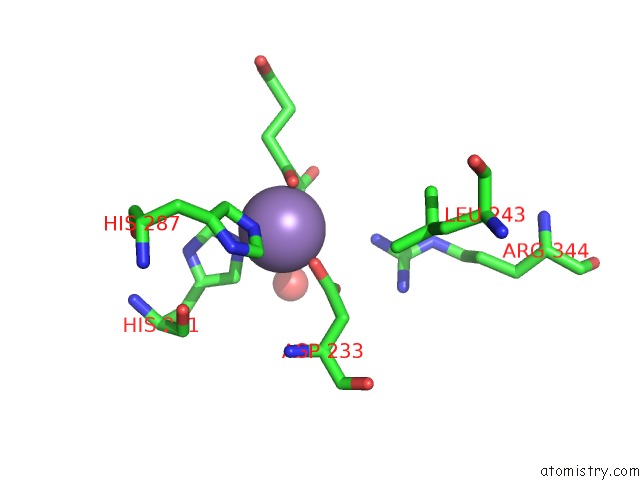
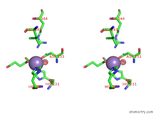
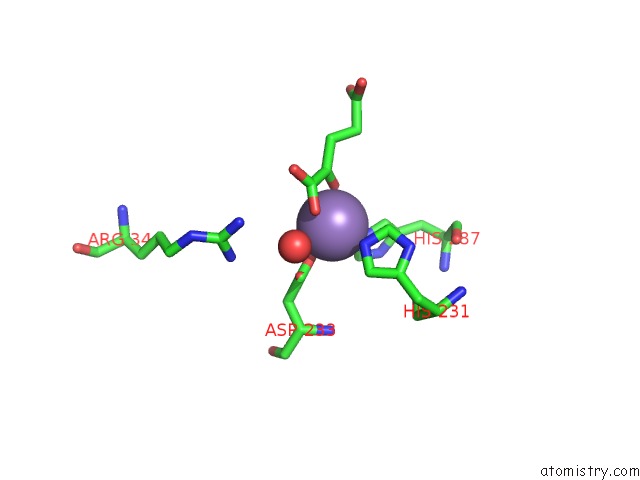
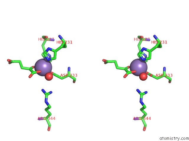
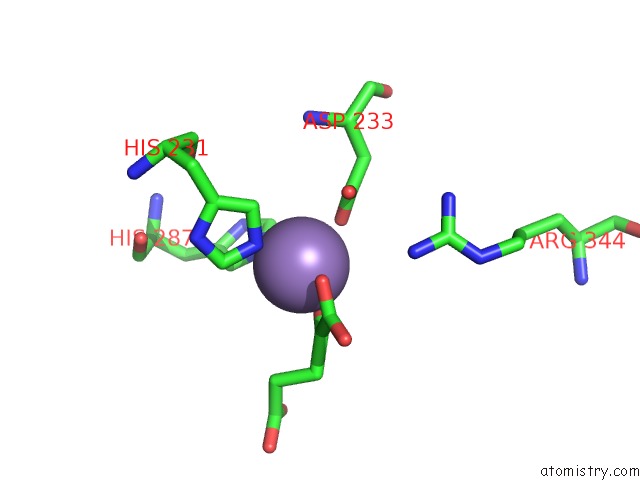
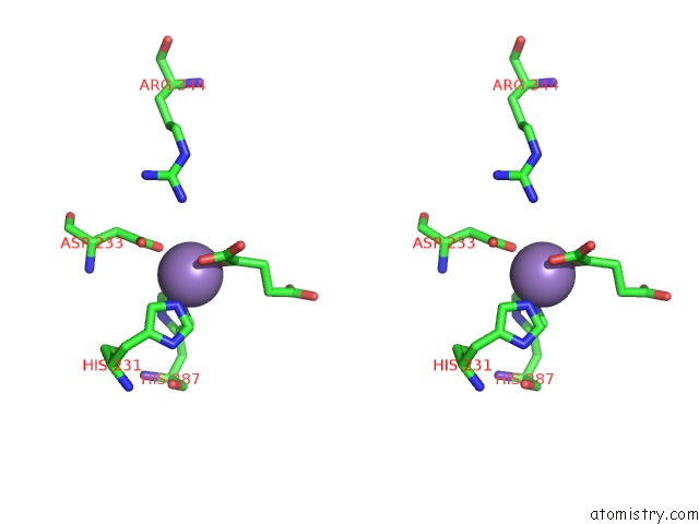
Manganese binding site 1 out of 8 in 6ie2
Go back to
Manganese binding site 1 out
of 8 in the Crystal Structure of Methyladenine Demethylase
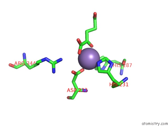
Mono view
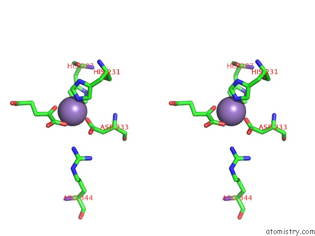
Stereo pair view
Mono view
Stereo pair view
A full contact list of Manganese with other atoms in the Mn binding
site number 1 of Crystal Structure of Methyladenine Demethylase within 5.0Å range:
|
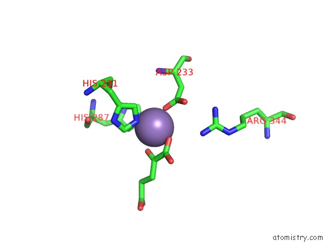
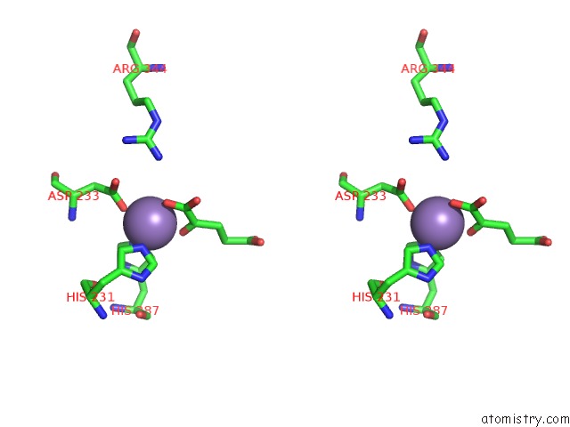
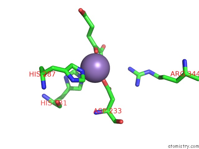
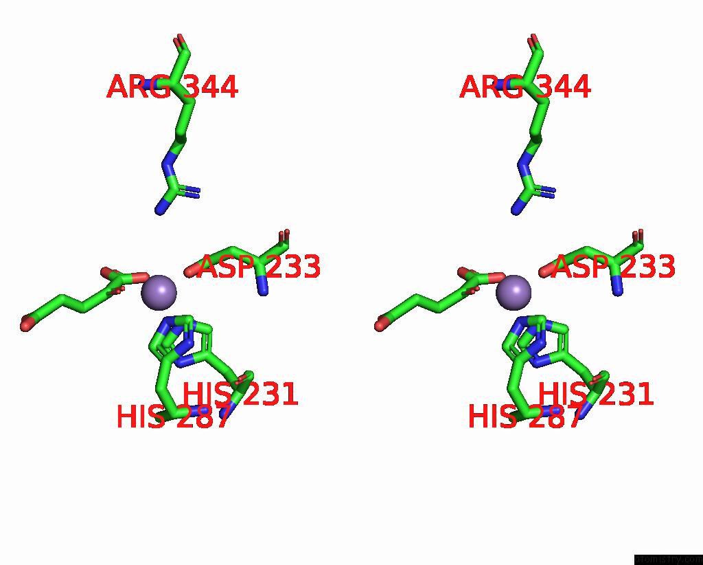
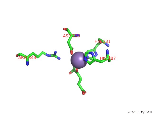
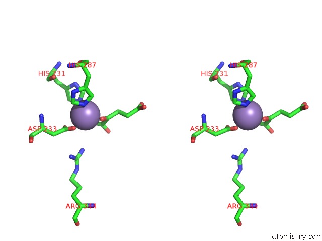
Manganese binding site 2 out of 8 in 6ie2
Go back to
Manganese binding site 2 out
of 8 in the Crystal Structure of Methyladenine Demethylase
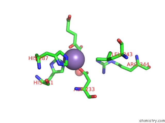
Mono view
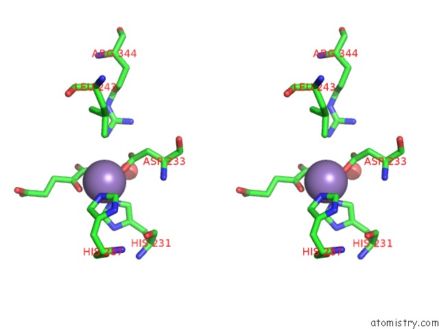
Stereo pair view
Mono view
Stereo pair view
A full contact list of Manganese with other atoms in the Mn binding
site number 2 of Crystal Structure of Methyladenine Demethylase within 5.0Å range:
|
Manganese binding site 3 out of 8 in 6ie2
Go back to
Manganese binding site 3 out
of 8 in the Crystal Structure of Methyladenine Demethylase
Mono view
Stereo pair view
Mono view
Stereo pair view
A full contact list of Manganese with other atoms in the Mn binding
site number 3 of Crystal Structure of Methyladenine Demethylase within 5.0Å range:
|
Manganese binding site 4 out of 8 in 6ie2
Go back to
Manganese binding site 4 out
of 8 in the Crystal Structure of Methyladenine Demethylase
Mono view
Stereo pair view
Mono view
Stereo pair view
A full contact list of Manganese with other atoms in the Mn binding
site number 4 of Crystal Structure of Methyladenine Demethylase within 5.0Å range:
|
Manganese binding site 5 out of 8 in 6ie2
Go back to
Manganese binding site 5 out
of 8 in the Crystal Structure of Methyladenine Demethylase
Mono view
Stereo pair view
Mono view
Stereo pair view
A full contact list of Manganese with other atoms in the Mn binding
site number 5 of Crystal Structure of Methyladenine Demethylase within 5.0Å range:
|
Manganese binding site 6 out of 8 in 6ie2
Go back to
Manganese binding site 6 out
of 8 in the Crystal Structure of Methyladenine Demethylase
Mono view
Stereo pair view
Mono view
Stereo pair view
A full contact list of Manganese with other atoms in the Mn binding
site number 6 of Crystal Structure of Methyladenine Demethylase within 5.0Å range:
|
Manganese binding site 7 out of 8 in 6ie2
Go back to
Manganese binding site 7 out
of 8 in the Crystal Structure of Methyladenine Demethylase
Mono view
Stereo pair view
Mono view
Stereo pair view
A full contact list of Manganese with other atoms in the Mn binding
site number 7 of Crystal Structure of Methyladenine Demethylase within 5.0Å range:
|
Manganese binding site 8 out of 8 in 6ie2
Go back to
Manganese binding site 8 out
of 8 in the Crystal Structure of Methyladenine Demethylase
Mono view
Stereo pair view
Mono view
Stereo pair view
A full contact list of Manganese with other atoms in the Mn binding
site number 8 of Crystal Structure of Methyladenine Demethylase within 5.0Å range:
|
Reference:
L.F.Tian,
Q.Tang,
Z.Z.Chen,
X.X.Yan.
Crystal Structure of Methyladenine Demethylase To Be Published.
Page generated: Sun Oct 6 04:52:57 2024
Last articles
Cl in 3DN1Cl in 3DN6
Cl in 3DN8
Cl in 3DN0
Cl in 3DMZ
Cl in 3DMX
Cl in 3DMV
Cl in 3DMJ
Cl in 3DM2
Cl in 3DLP