Manganese »
PDB 6fxt-6hzn »
6h5a »
Manganese in PDB 6h5a: Crystal Structure of Mycobacterium Tuberculosis Phosphatidylinositol Phosphate Synthase (PGSA1) in Complex with Manganese and Citrate
Enzymatic activity of Crystal Structure of Mycobacterium Tuberculosis Phosphatidylinositol Phosphate Synthase (PGSA1) in Complex with Manganese and Citrate
All present enzymatic activity of Crystal Structure of Mycobacterium Tuberculosis Phosphatidylinositol Phosphate Synthase (PGSA1) in Complex with Manganese and Citrate:
2.7.8.11;
2.7.8.11;
Protein crystallography data
The structure of Crystal Structure of Mycobacterium Tuberculosis Phosphatidylinositol Phosphate Synthase (PGSA1) in Complex with Manganese and Citrate, PDB code: 6h5a
was solved by
K.Grave,
M.Hogbom,
with X-Ray Crystallography technique. A brief refinement statistics is given in the table below:
| Resolution Low / High (Å) | 45.70 / 1.88 |
| Space group | P 21 21 21 |
| Cell size a, b, c (Å), α, β, γ (°) | 68.067, 77.941, 100.823, 90.00, 90.00, 90.00 |
| R / Rfree (%) | 20.4 / 23 |
Manganese Binding Sites:
The binding sites of Manganese atom in the Crystal Structure of Mycobacterium Tuberculosis Phosphatidylinositol Phosphate Synthase (PGSA1) in Complex with Manganese and Citrate
(pdb code 6h5a). This binding sites where shown within
5.0 Angstroms radius around Manganese atom.
In total 7 binding sites of Manganese where determined in the Crystal Structure of Mycobacterium Tuberculosis Phosphatidylinositol Phosphate Synthase (PGSA1) in Complex with Manganese and Citrate, PDB code: 6h5a:
Jump to Manganese binding site number: 1; 2; 3; 4; 5; 6; 7;
In total 7 binding sites of Manganese where determined in the Crystal Structure of Mycobacterium Tuberculosis Phosphatidylinositol Phosphate Synthase (PGSA1) in Complex with Manganese and Citrate, PDB code: 6h5a:
Jump to Manganese binding site number: 1; 2; 3; 4; 5; 6; 7;
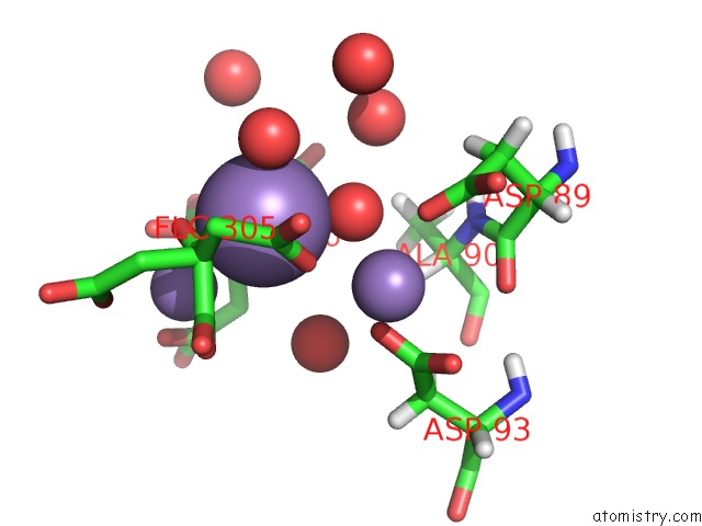
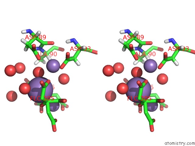
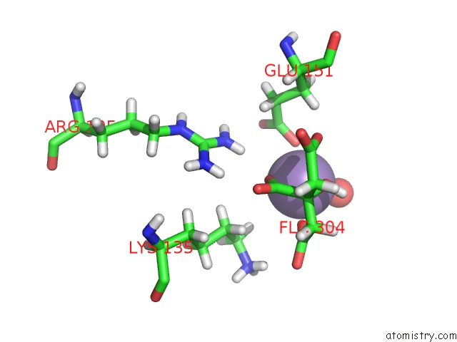
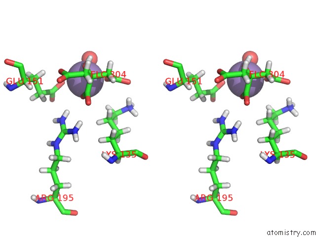
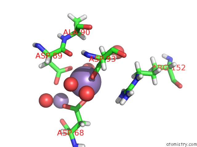
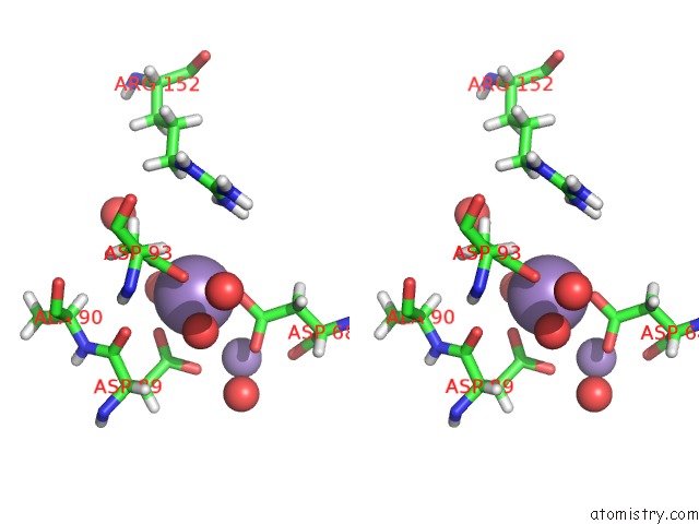
Manganese binding site 1 out of 7 in 6h5a
Go back to
Manganese binding site 1 out
of 7 in the Crystal Structure of Mycobacterium Tuberculosis Phosphatidylinositol Phosphate Synthase (PGSA1) in Complex with Manganese and Citrate
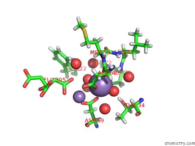
Mono view
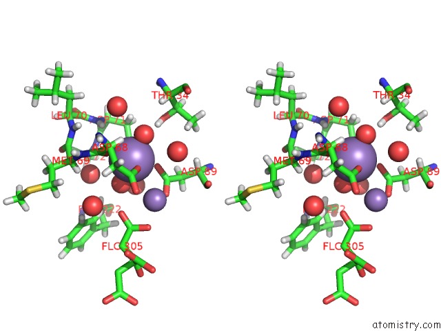
Stereo pair view
Mono view
Stereo pair view
A full contact list of Manganese with other atoms in the Mn binding
site number 1 of Crystal Structure of Mycobacterium Tuberculosis Phosphatidylinositol Phosphate Synthase (PGSA1) in Complex with Manganese and Citrate within 5.0Å range:
|
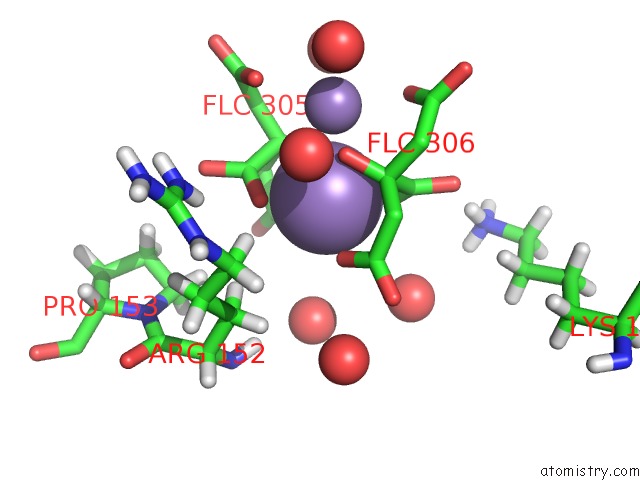
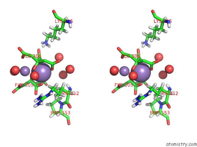
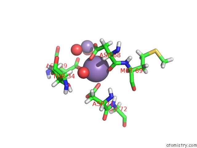
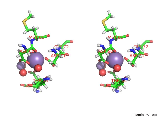
Manganese binding site 2 out of 7 in 6h5a
Go back to
Manganese binding site 2 out
of 7 in the Crystal Structure of Mycobacterium Tuberculosis Phosphatidylinositol Phosphate Synthase (PGSA1) in Complex with Manganese and Citrate
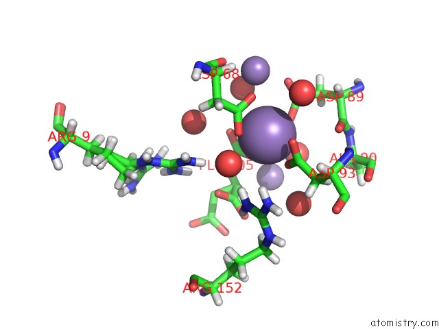
Mono view
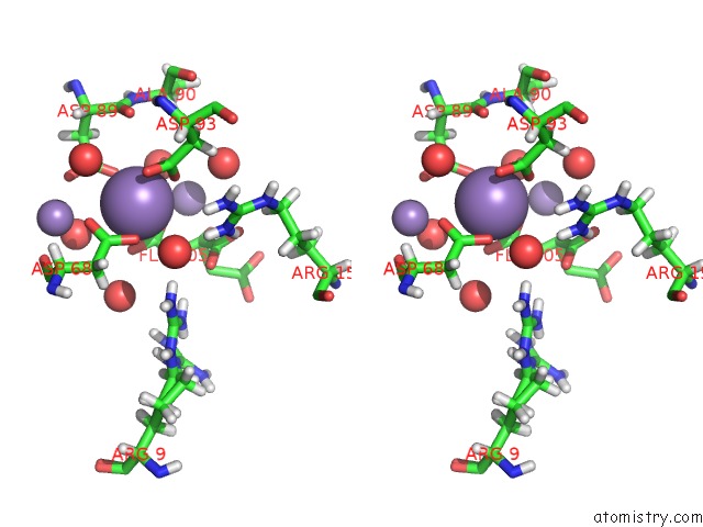
Stereo pair view
Mono view
Stereo pair view
A full contact list of Manganese with other atoms in the Mn binding
site number 2 of Crystal Structure of Mycobacterium Tuberculosis Phosphatidylinositol Phosphate Synthase (PGSA1) in Complex with Manganese and Citrate within 5.0Å range:
|
Manganese binding site 3 out of 7 in 6h5a
Go back to
Manganese binding site 3 out
of 7 in the Crystal Structure of Mycobacterium Tuberculosis Phosphatidylinositol Phosphate Synthase (PGSA1) in Complex with Manganese and Citrate
Mono view
Stereo pair view
Mono view
Stereo pair view
A full contact list of Manganese with other atoms in the Mn binding
site number 3 of Crystal Structure of Mycobacterium Tuberculosis Phosphatidylinositol Phosphate Synthase (PGSA1) in Complex with Manganese and Citrate within 5.0Å range:
|
Manganese binding site 4 out of 7 in 6h5a
Go back to
Manganese binding site 4 out
of 7 in the Crystal Structure of Mycobacterium Tuberculosis Phosphatidylinositol Phosphate Synthase (PGSA1) in Complex with Manganese and Citrate
Mono view
Stereo pair view
Mono view
Stereo pair view
A full contact list of Manganese with other atoms in the Mn binding
site number 4 of Crystal Structure of Mycobacterium Tuberculosis Phosphatidylinositol Phosphate Synthase (PGSA1) in Complex with Manganese and Citrate within 5.0Å range:
|
Manganese binding site 5 out of 7 in 6h5a
Go back to
Manganese binding site 5 out
of 7 in the Crystal Structure of Mycobacterium Tuberculosis Phosphatidylinositol Phosphate Synthase (PGSA1) in Complex with Manganese and Citrate
Mono view
Stereo pair view
Mono view
Stereo pair view
A full contact list of Manganese with other atoms in the Mn binding
site number 5 of Crystal Structure of Mycobacterium Tuberculosis Phosphatidylinositol Phosphate Synthase (PGSA1) in Complex with Manganese and Citrate within 5.0Å range:
|
Manganese binding site 6 out of 7 in 6h5a
Go back to
Manganese binding site 6 out
of 7 in the Crystal Structure of Mycobacterium Tuberculosis Phosphatidylinositol Phosphate Synthase (PGSA1) in Complex with Manganese and Citrate
Mono view
Stereo pair view
Mono view
Stereo pair view
A full contact list of Manganese with other atoms in the Mn binding
site number 6 of Crystal Structure of Mycobacterium Tuberculosis Phosphatidylinositol Phosphate Synthase (PGSA1) in Complex with Manganese and Citrate within 5.0Å range:
|
Manganese binding site 7 out of 7 in 6h5a
Go back to
Manganese binding site 7 out
of 7 in the Crystal Structure of Mycobacterium Tuberculosis Phosphatidylinositol Phosphate Synthase (PGSA1) in Complex with Manganese and Citrate
Mono view
Stereo pair view
Mono view
Stereo pair view
A full contact list of Manganese with other atoms in the Mn binding
site number 7 of Crystal Structure of Mycobacterium Tuberculosis Phosphatidylinositol Phosphate Synthase (PGSA1) in Complex with Manganese and Citrate within 5.0Å range:
|
Reference:
K.Grave,
M.D.Bennett,
M.Hogbom.
Structure Ofmycobacterium Tuberculosisphosphatidylinositol Phosphate Synthase Reveals Mechanism of Substrate Binding and Metal Catalysis. Commun Biol V. 2 175 2019.
ISSN: ESSN 2399-3642
PubMed: 31098408
DOI: 10.1038/S42003-019-0427-1
Page generated: Sun Oct 6 04:50:17 2024
ISSN: ESSN 2399-3642
PubMed: 31098408
DOI: 10.1038/S42003-019-0427-1
Last articles
Zn in 9J0NZn in 9J0O
Zn in 9J0P
Zn in 9FJX
Zn in 9EKB
Zn in 9C0F
Zn in 9CAH
Zn in 9CH0
Zn in 9CH3
Zn in 9CH1