Manganese »
PDB 6f4q-6fxr »
6fm4 »
Manganese in PDB 6fm4: The Crystal Structure of S. Aureus Gyrase Complex with Id-130 and Dna
Enzymatic activity of The Crystal Structure of S. Aureus Gyrase Complex with Id-130 and Dna
All present enzymatic activity of The Crystal Structure of S. Aureus Gyrase Complex with Id-130 and Dna:
5.99.1.3;
5.99.1.3;
Protein crystallography data
The structure of The Crystal Structure of S. Aureus Gyrase Complex with Id-130 and Dna, PDB code: 6fm4
was solved by
R.Ombrato,
B.Garofalo,
G.Mangano,
F.Mancini,
with X-Ray Crystallography technique. A brief refinement statistics is given in the table below:
| Resolution Low / High (Å) | 43.05 / 2.70 |
| Space group | P 61 |
| Cell size a, b, c (Å), α, β, γ (°) | 92.951, 92.951, 407.030, 90.00, 90.00, 120.00 |
| R / Rfree (%) | 22.5 / 30 |
Manganese Binding Sites:
The binding sites of Manganese atom in the The Crystal Structure of S. Aureus Gyrase Complex with Id-130 and Dna
(pdb code 6fm4). This binding sites where shown within
5.0 Angstroms radius around Manganese atom.
In total 3 binding sites of Manganese where determined in the The Crystal Structure of S. Aureus Gyrase Complex with Id-130 and Dna, PDB code: 6fm4:
Jump to Manganese binding site number: 1; 2; 3;
In total 3 binding sites of Manganese where determined in the The Crystal Structure of S. Aureus Gyrase Complex with Id-130 and Dna, PDB code: 6fm4:
Jump to Manganese binding site number: 1; 2; 3;
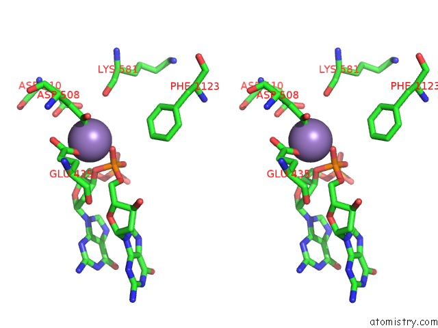
Manganese binding site 1 out of 3 in 6fm4
Go back to
Manganese binding site 1 out
of 3 in the The Crystal Structure of S. Aureus Gyrase Complex with Id-130 and Dna

Mono view
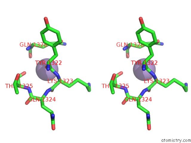
Stereo pair view
Mono view
Stereo pair view
A full contact list of Manganese with other atoms in the Mn binding
site number 1 of The Crystal Structure of S. Aureus Gyrase Complex with Id-130 and Dna within 5.0Å range:
|
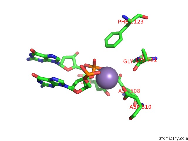
Manganese binding site 2 out of 3 in 6fm4
Go back to
Manganese binding site 2 out
of 3 in the The Crystal Structure of S. Aureus Gyrase Complex with Id-130 and Dna
Mono view
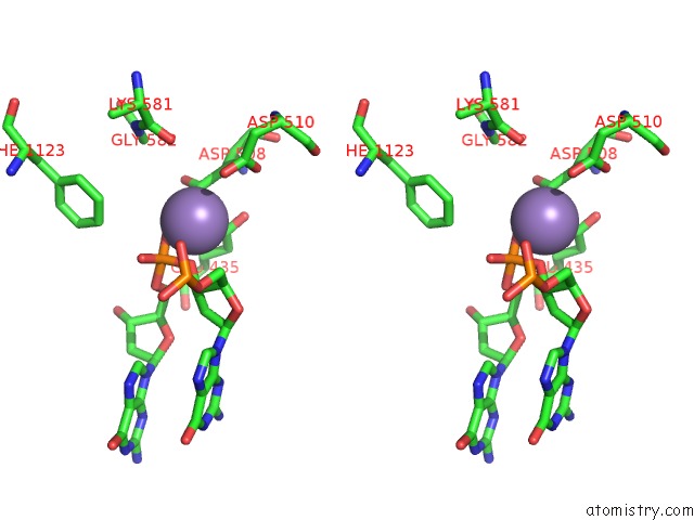
Stereo pair view
Mono view
Stereo pair view
A full contact list of Manganese with other atoms in the Mn binding
site number 2 of The Crystal Structure of S. Aureus Gyrase Complex with Id-130 and Dna within 5.0Å range:
|
Manganese binding site 3 out of 3 in 6fm4
Go back to
Manganese binding site 3 out
of 3 in the The Crystal Structure of S. Aureus Gyrase Complex with Id-130 and Dna
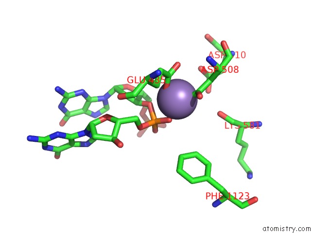
Mono view
Stereo pair view
Mono view
Stereo pair view
A full contact list of Manganese with other atoms in the Mn binding
site number 3 of The Crystal Structure of S. Aureus Gyrase Complex with Id-130 and Dna within 5.0Å range:
|
Reference:
G.Magaro,
F.Prati,
B.Garofalo,
G.Corso,
G.Furlotti,
C.Apicella,
G.Mangano,
N.D'atanasio,
D.Robinson,
F.P.Di Giorgio,
R.Ombrato.
Virtual Screening Approach and Investigation of Structure-Activity Relationships to Discover Novel Bacterial Topoisomerase Inhibitors Targeting Gram-Positive and Gram-Negative Pathogens. J.Med.Chem. V. 62 7445 2019.
ISSN: ISSN 0022-2623
PubMed: 31276392
DOI: 10.1021/ACS.JMEDCHEM.9B00394
Page generated: Sun Oct 6 04:37:15 2024
ISSN: ISSN 0022-2623
PubMed: 31276392
DOI: 10.1021/ACS.JMEDCHEM.9B00394
Last articles
Zn in 9J0NZn in 9J0O
Zn in 9J0P
Zn in 9FJX
Zn in 9EKB
Zn in 9C0F
Zn in 9CAH
Zn in 9CH0
Zn in 9CH3
Zn in 9CH1