Manganese »
PDB 6e4q-6f4p »
6ev5 »
Manganese in PDB 6ev5: Crystal Structure of E282Q A. Niger FDC1 with Prfmn in the Hydroxylated Form
Enzymatic activity of Crystal Structure of E282Q A. Niger FDC1 with Prfmn in the Hydroxylated Form
All present enzymatic activity of Crystal Structure of E282Q A. Niger FDC1 with Prfmn in the Hydroxylated Form:
4.1.1.102;
4.1.1.102;
Protein crystallography data
The structure of Crystal Structure of E282Q A. Niger FDC1 with Prfmn in the Hydroxylated Form, PDB code: 6ev5
was solved by
S.S.Bailey,
D.Leys,
K.A.P.Payne,
with X-Ray Crystallography technique. A brief refinement statistics is given in the table below:
| Resolution Low / High (Å) | 38.30 / 1.28 |
| Space group | P 21 21 2 |
| Cell size a, b, c (Å), α, β, γ (°) | 96.020, 63.510, 87.620, 90.00, 90.00, 90.00 |
| R / Rfree (%) | 16.6 / 19 |
Other elements in 6ev5:
The structure of Crystal Structure of E282Q A. Niger FDC1 with Prfmn in the Hydroxylated Form also contains other interesting chemical elements:
| Potassium | (K) | 2 atoms |
Manganese Binding Sites:
The binding sites of Manganese atom in the Crystal Structure of E282Q A. Niger FDC1 with Prfmn in the Hydroxylated Form
(pdb code 6ev5). This binding sites where shown within
5.0 Angstroms radius around Manganese atom.
In total only one binding site of Manganese was determined in the Crystal Structure of E282Q A. Niger FDC1 with Prfmn in the Hydroxylated Form, PDB code: 6ev5:
In total only one binding site of Manganese was determined in the Crystal Structure of E282Q A. Niger FDC1 with Prfmn in the Hydroxylated Form, PDB code: 6ev5:
Manganese binding site 1 out of 1 in 6ev5
Go back to
Manganese binding site 1 out
of 1 in the Crystal Structure of E282Q A. Niger FDC1 with Prfmn in the Hydroxylated Form
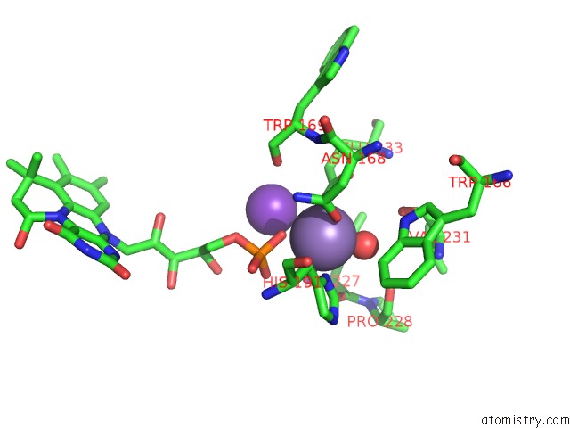
Mono view
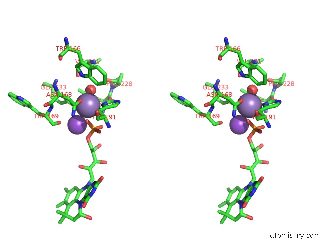
Stereo pair view
Mono view
Stereo pair view
A full contact list of Manganese with other atoms in the Mn binding
site number 1 of Crystal Structure of E282Q A. Niger FDC1 with Prfmn in the Hydroxylated Form within 5.0Å range:
|
Reference:
S.S.Bailey,
K.A.P.Payne,
K.Fisher,
S.A.Marshall,
M.J.Cliff,
R.Spiess,
D.A.Parker,
S.E.J.Rigby,
D.Leys.
The Role of Conserved Residues in Fdc Decarboxylase in Prenylated Flavin Mononucleotide Oxidative Maturation, Cofactor Isomerization, and Catalysis. J. Biol. Chem. V. 293 2272 2018.
ISSN: ESSN 1083-351X
PubMed: 29259125
DOI: 10.1074/JBC.RA117.000881
Page generated: Sun Oct 6 04:20:40 2024
ISSN: ESSN 1083-351X
PubMed: 29259125
DOI: 10.1074/JBC.RA117.000881
Last articles
Zn in 9J0NZn in 9J0O
Zn in 9J0P
Zn in 9FJX
Zn in 9EKB
Zn in 9C0F
Zn in 9CAH
Zn in 9CH0
Zn in 9CH3
Zn in 9CH1