Manganese »
PDB 6e4q-6f4p »
6egs »
Manganese in PDB 6egs: Crystal Structure of the Galnac-T2 F104S Mutant in Complex with Udp- Galnac
Enzymatic activity of Crystal Structure of the Galnac-T2 F104S Mutant in Complex with Udp- Galnac
All present enzymatic activity of Crystal Structure of the Galnac-T2 F104S Mutant in Complex with Udp- Galnac:
2.4.1.41;
2.4.1.41;
Protein crystallography data
The structure of Crystal Structure of the Galnac-T2 F104S Mutant in Complex with Udp- Galnac, PDB code: 6egs
was solved by
M.De Las Rivas,
H.Coelho,
A.Diniz,
E.Lira-Navarrete,
J.Jimenez-Barbero,
K.T.Schjoldager,
E.P.Bennett,
S.Y.Vakhrushev,
H.Clausen,
F.Corzana,
F.Marcelo,
R.Hurtado-Guerrero,
with X-Ray Crystallography technique. A brief refinement statistics is given in the table below:
| Resolution Low / High (Å) | 154.04 / 2.70 |
| Space group | P 41 21 2 |
| Cell size a, b, c (Å), α, β, γ (°) | 154.036, 154.036, 109.823, 90.00, 90.00, 90.00 |
| R / Rfree (%) | 20.6 / 22.1 |
Manganese Binding Sites:
The binding sites of Manganese atom in the Crystal Structure of the Galnac-T2 F104S Mutant in Complex with Udp- Galnac
(pdb code 6egs). This binding sites where shown within
5.0 Angstroms radius around Manganese atom.
In total 2 binding sites of Manganese where determined in the Crystal Structure of the Galnac-T2 F104S Mutant in Complex with Udp- Galnac, PDB code: 6egs:
Jump to Manganese binding site number: 1; 2;
In total 2 binding sites of Manganese where determined in the Crystal Structure of the Galnac-T2 F104S Mutant in Complex with Udp- Galnac, PDB code: 6egs:
Jump to Manganese binding site number: 1; 2;
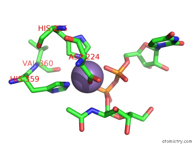
Manganese binding site 1 out of 2 in 6egs
Go back to
Manganese binding site 1 out
of 2 in the Crystal Structure of the Galnac-T2 F104S Mutant in Complex with Udp- Galnac
Mono view
Stereo pair view
Mono view
Stereo pair view
A full contact list of Manganese with other atoms in the Mn binding
site number 1 of Crystal Structure of the Galnac-T2 F104S Mutant in Complex with Udp- Galnac within 5.0Å range:
|
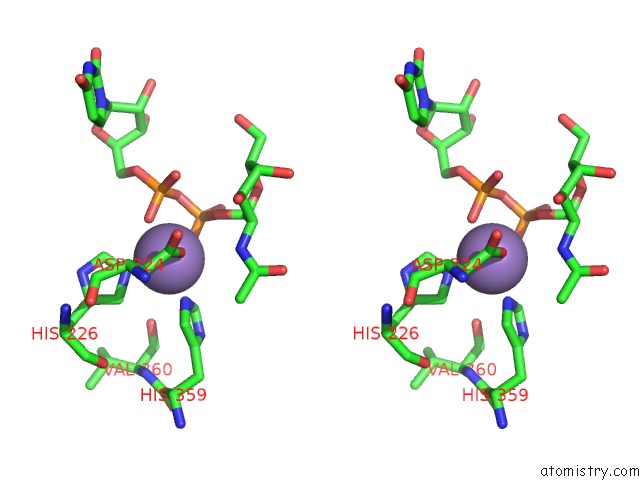
Manganese binding site 2 out of 2 in 6egs
Go back to
Manganese binding site 2 out
of 2 in the Crystal Structure of the Galnac-T2 F104S Mutant in Complex with Udp- Galnac

Mono view

Stereo pair view
Mono view
Stereo pair view
A full contact list of Manganese with other atoms in the Mn binding
site number 2 of Crystal Structure of the Galnac-T2 F104S Mutant in Complex with Udp- Galnac within 5.0Å range:
|
Reference:
M.De Las Rivas,
H.Coelho,
A.Diniz,
E.Lira-Navarrete,
I.Companon,
J.Jimenez-Barbero,
K.T.Schjoldager,
E.P.Bennett,
S.Y.Vakhrushev,
H.Clausen,
F.Corzana,
F.Marcelo,
R.Hurtado-Guerrero.
Structural Analysis of A Galnac-T2 Mutant Reveals An Induced-Fit Catalytic Mechanism For Galnac-Ts. Chemistry V. 24 8382 2018.
ISSN: ISSN 1521-3765
PubMed: 29601100
DOI: 10.1002/CHEM.201800701
Page generated: Sun Oct 6 04:18:58 2024
ISSN: ISSN 1521-3765
PubMed: 29601100
DOI: 10.1002/CHEM.201800701
Last articles
Zn in 9J0NZn in 9J0O
Zn in 9J0P
Zn in 9FJX
Zn in 9EKB
Zn in 9C0F
Zn in 9CAH
Zn in 9CH0
Zn in 9CH3
Zn in 9CH1