Manganese »
PDB 6bh5-6dk4 »
6d0p »
Manganese in PDB 6d0p: 1.88 Angstrom Resolution Crystal Structure of Quercetin 2,3- Dioxygenase From Acinetobacter Baumannii
Protein crystallography data
The structure of 1.88 Angstrom Resolution Crystal Structure of Quercetin 2,3- Dioxygenase From Acinetobacter Baumannii, PDB code: 6d0p
was solved by
G.Minasov,
L.Shuvalova,
J.S.Brunzelle,
I.Dubrovska,
O.Kiryukhina,
M.Endres,
W.F.Anderson,
K.J.F.Satchell,
A.Joachimiak,
Center Forstructural Genomics Of Infectious Diseases (Csgid),
with X-Ray Crystallography technique. A brief refinement statistics is given in the table below:
| Resolution Low / High (Å) | 29.81 / 1.88 |
| Space group | C 1 2 1 |
| Cell size a, b, c (Å), α, β, γ (°) | 95.481, 131.977, 123.987, 90.00, 103.58, 90.00 |
| R / Rfree (%) | 15.8 / 19 |
Other elements in 6d0p:
The structure of 1.88 Angstrom Resolution Crystal Structure of Quercetin 2,3- Dioxygenase From Acinetobacter Baumannii also contains other interesting chemical elements:
| Magnesium | (Mg) | 2 atoms |
| Chlorine | (Cl) | 2 atoms |
Manganese Binding Sites:
The binding sites of Manganese atom in the 1.88 Angstrom Resolution Crystal Structure of Quercetin 2,3- Dioxygenase From Acinetobacter Baumannii
(pdb code 6d0p). This binding sites where shown within
5.0 Angstroms radius around Manganese atom.
In total 4 binding sites of Manganese where determined in the 1.88 Angstrom Resolution Crystal Structure of Quercetin 2,3- Dioxygenase From Acinetobacter Baumannii, PDB code: 6d0p:
Jump to Manganese binding site number: 1; 2; 3; 4;
In total 4 binding sites of Manganese where determined in the 1.88 Angstrom Resolution Crystal Structure of Quercetin 2,3- Dioxygenase From Acinetobacter Baumannii, PDB code: 6d0p:
Jump to Manganese binding site number: 1; 2; 3; 4;
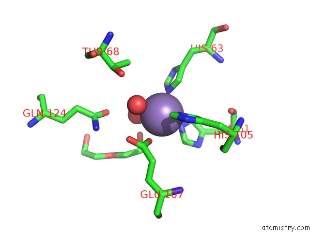
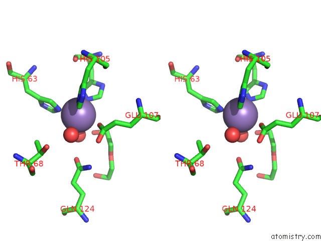
Manganese binding site 1 out of 4 in 6d0p
Go back to
Manganese binding site 1 out
of 4 in the 1.88 Angstrom Resolution Crystal Structure of Quercetin 2,3- Dioxygenase From Acinetobacter Baumannii
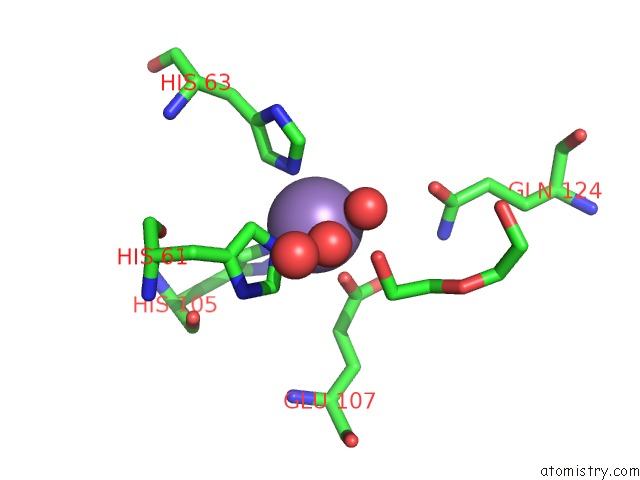
Mono view
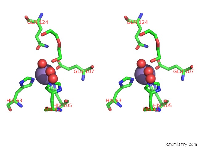
Stereo pair view
Mono view
Stereo pair view
A full contact list of Manganese with other atoms in the Mn binding
site number 1 of 1.88 Angstrom Resolution Crystal Structure of Quercetin 2,3- Dioxygenase From Acinetobacter Baumannii within 5.0Å range:
|
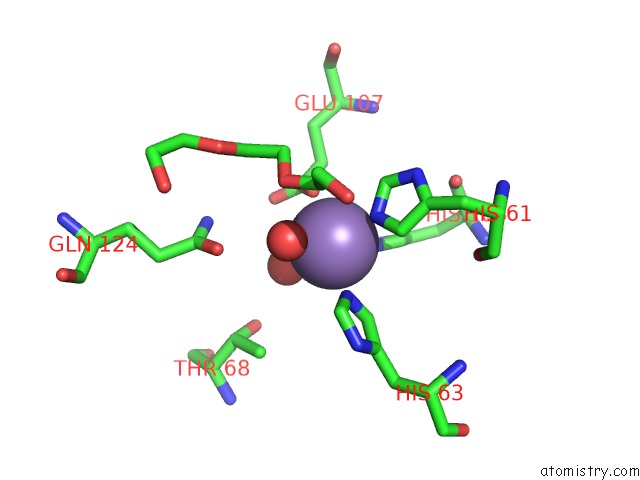
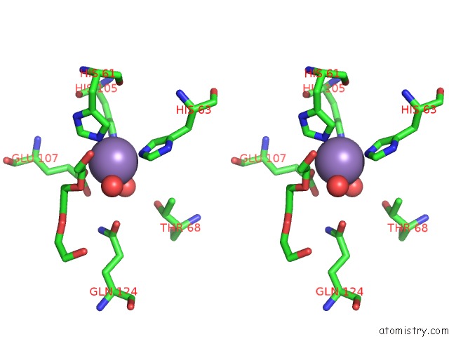
Manganese binding site 2 out of 4 in 6d0p
Go back to
Manganese binding site 2 out
of 4 in the 1.88 Angstrom Resolution Crystal Structure of Quercetin 2,3- Dioxygenase From Acinetobacter Baumannii
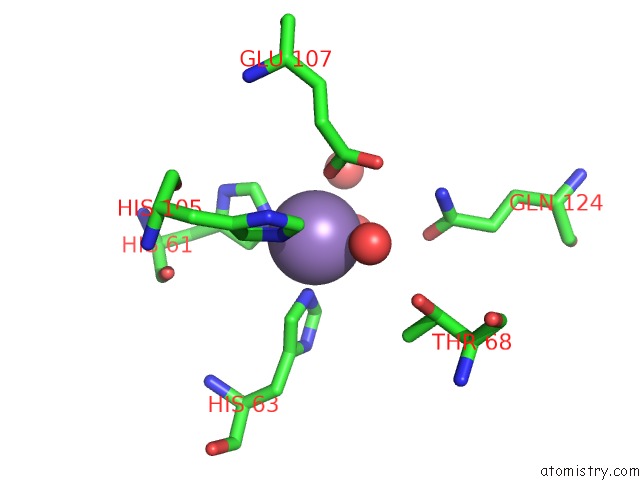
Mono view
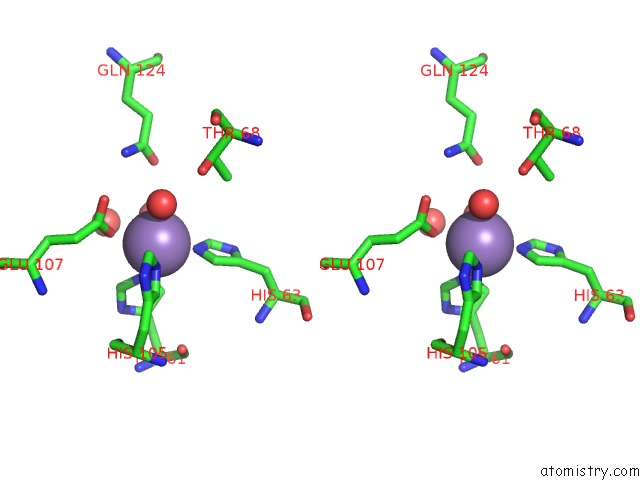
Stereo pair view
Mono view
Stereo pair view
A full contact list of Manganese with other atoms in the Mn binding
site number 2 of 1.88 Angstrom Resolution Crystal Structure of Quercetin 2,3- Dioxygenase From Acinetobacter Baumannii within 5.0Å range:
|
Manganese binding site 3 out of 4 in 6d0p
Go back to
Manganese binding site 3 out
of 4 in the 1.88 Angstrom Resolution Crystal Structure of Quercetin 2,3- Dioxygenase From Acinetobacter Baumannii
Mono view
Stereo pair view
Mono view
Stereo pair view
A full contact list of Manganese with other atoms in the Mn binding
site number 3 of 1.88 Angstrom Resolution Crystal Structure of Quercetin 2,3- Dioxygenase From Acinetobacter Baumannii within 5.0Å range:
|
Manganese binding site 4 out of 4 in 6d0p
Go back to
Manganese binding site 4 out
of 4 in the 1.88 Angstrom Resolution Crystal Structure of Quercetin 2,3- Dioxygenase From Acinetobacter Baumannii
Mono view
Stereo pair view
Mono view
Stereo pair view
A full contact list of Manganese with other atoms in the Mn binding
site number 4 of 1.88 Angstrom Resolution Crystal Structure of Quercetin 2,3- Dioxygenase From Acinetobacter Baumannii within 5.0Å range:
|
Reference:
G.Minasov,
L.Shuvalova,
J.S.Brunzelle,
I.Dubrovska,
O.Kiryukhina,
M.Endres,
W.F.Anderson,
K.J.F.Satchell,
A.Joachimiak.
1.88 Angstrom Resolution Crystal Structure of Quercetin 2,3-Dioxygenase From Acinetobacter Baumannii To Be Published.
Page generated: Sun Oct 6 04:02:34 2024
Last articles
F in 4HN4F in 4HJX
F in 4HLH
F in 4HL4
F in 4HIQ
F in 4HHZ
F in 4HHY
F in 4HGT
F in 4HEJ
F in 4HGS