Manganese »
PDB 6bh5-6dk4 »
6cwq »
Manganese in PDB 6cwq: X-Ray Crystal Structure of Flavobacterium Johnsoniae Dimanganese(II) Ribonucleotide Reductase Beta Subunit (As-Isolated)
Protein crystallography data
The structure of X-Ray Crystal Structure of Flavobacterium Johnsoniae Dimanganese(II) Ribonucleotide Reductase Beta Subunit (As-Isolated), PDB code: 6cwq
was solved by
H.R.Rose,
A.O.Maggiolo,
A.K.Boal,
with X-Ray Crystallography technique. A brief refinement statistics is given in the table below:
| Resolution Low / High (Å) | 72.63 / 1.90 |
| Space group | P 1 |
| Cell size a, b, c (Å), α, β, γ (°) | 48.356, 50.816, 79.285, 106.15, 105.03, 95.75 |
| R / Rfree (%) | 19.2 / 21.2 |
Other elements in 6cwq:
The structure of X-Ray Crystal Structure of Flavobacterium Johnsoniae Dimanganese(II) Ribonucleotide Reductase Beta Subunit (As-Isolated) also contains other interesting chemical elements:
| Magnesium | (Mg) | 2 atoms |
Manganese Binding Sites:
The binding sites of Manganese atom in the X-Ray Crystal Structure of Flavobacterium Johnsoniae Dimanganese(II) Ribonucleotide Reductase Beta Subunit (As-Isolated)
(pdb code 6cwq). This binding sites where shown within
5.0 Angstroms radius around Manganese atom.
In total 4 binding sites of Manganese where determined in the X-Ray Crystal Structure of Flavobacterium Johnsoniae Dimanganese(II) Ribonucleotide Reductase Beta Subunit (As-Isolated), PDB code: 6cwq:
Jump to Manganese binding site number: 1; 2; 3; 4;
In total 4 binding sites of Manganese where determined in the X-Ray Crystal Structure of Flavobacterium Johnsoniae Dimanganese(II) Ribonucleotide Reductase Beta Subunit (As-Isolated), PDB code: 6cwq:
Jump to Manganese binding site number: 1; 2; 3; 4;
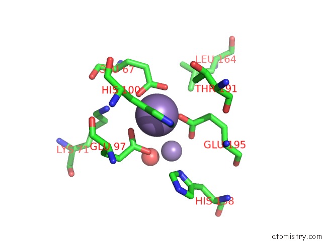
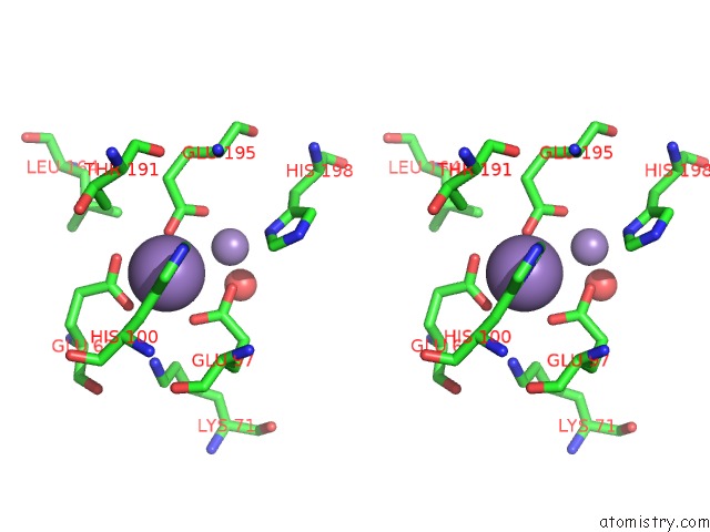
Manganese binding site 1 out of 4 in 6cwq
Go back to
Manganese binding site 1 out
of 4 in the X-Ray Crystal Structure of Flavobacterium Johnsoniae Dimanganese(II) Ribonucleotide Reductase Beta Subunit (As-Isolated)
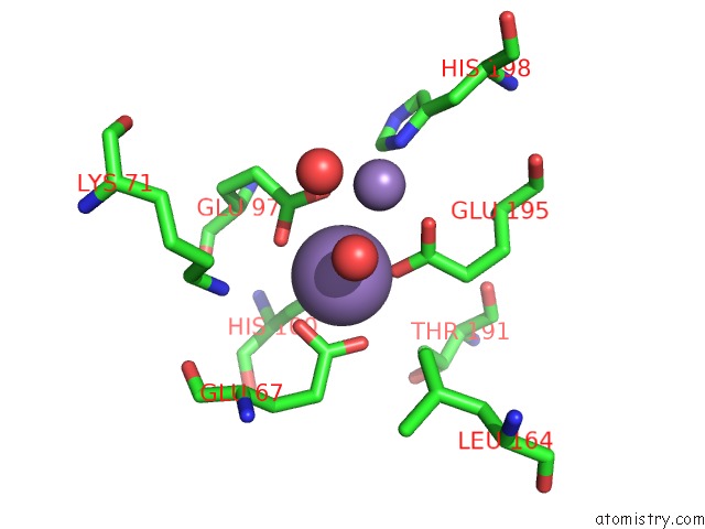
Mono view
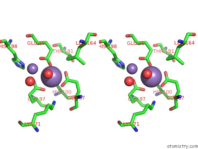
Stereo pair view
Mono view
Stereo pair view
A full contact list of Manganese with other atoms in the Mn binding
site number 1 of X-Ray Crystal Structure of Flavobacterium Johnsoniae Dimanganese(II) Ribonucleotide Reductase Beta Subunit (As-Isolated) within 5.0Å range:
|
Manganese binding site 2 out of 4 in 6cwq
Go back to
Manganese binding site 2 out
of 4 in the X-Ray Crystal Structure of Flavobacterium Johnsoniae Dimanganese(II) Ribonucleotide Reductase Beta Subunit (As-Isolated)
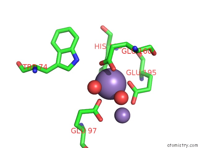
Mono view
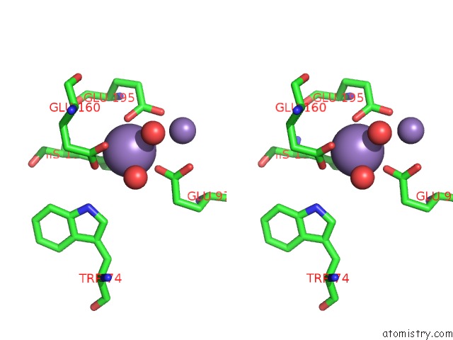
Stereo pair view
Mono view
Stereo pair view
A full contact list of Manganese with other atoms in the Mn binding
site number 2 of X-Ray Crystal Structure of Flavobacterium Johnsoniae Dimanganese(II) Ribonucleotide Reductase Beta Subunit (As-Isolated) within 5.0Å range:
|
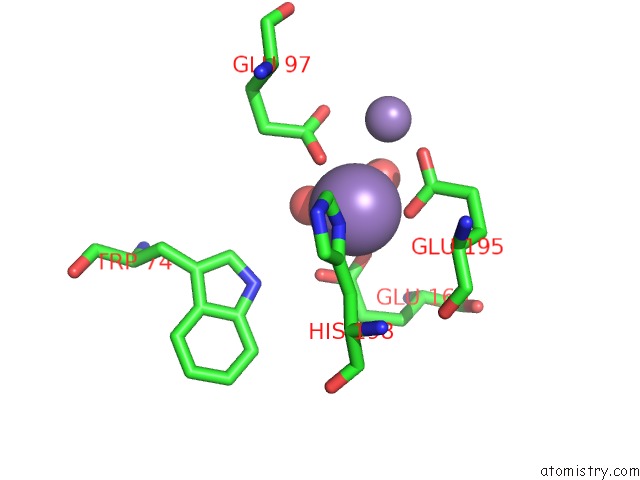
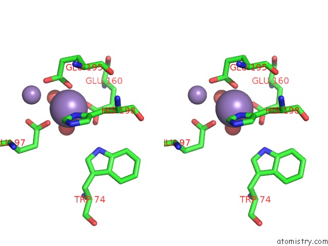
Manganese binding site 3 out of 4 in 6cwq
Go back to
Manganese binding site 3 out
of 4 in the X-Ray Crystal Structure of Flavobacterium Johnsoniae Dimanganese(II) Ribonucleotide Reductase Beta Subunit (As-Isolated)
Mono view
Stereo pair view
Mono view
Stereo pair view
A full contact list of Manganese with other atoms in the Mn binding
site number 3 of X-Ray Crystal Structure of Flavobacterium Johnsoniae Dimanganese(II) Ribonucleotide Reductase Beta Subunit (As-Isolated) within 5.0Å range:
|
Manganese binding site 4 out of 4 in 6cwq
Go back to
Manganese binding site 4 out
of 4 in the X-Ray Crystal Structure of Flavobacterium Johnsoniae Dimanganese(II) Ribonucleotide Reductase Beta Subunit (As-Isolated)
Mono view
Stereo pair view
Mono view
Stereo pair view
A full contact list of Manganese with other atoms in the Mn binding
site number 4 of X-Ray Crystal Structure of Flavobacterium Johnsoniae Dimanganese(II) Ribonucleotide Reductase Beta Subunit (As-Isolated) within 5.0Å range:
|
Reference:
H.R.Rose,
M.K.Ghosh,
A.O.Maggiolo,
C.J.Pollock,
E.J.Blaesi,
V.Hajj,
Y.Wei,
L.J.Rajakovich,
W.C.Chang,
Y.Han,
M.Hajj,
C.Krebs,
A.Silakov,
M.E.Pandelia,
J.M.Bollinger,
A.K.Boal.
Structural Basis For Superoxide Activation of Flavobacterium Johnsoniae Class I Ribonucleotide Reductase and For Radical Initiation By Its Dimanganese Cofactor. Biochemistry V. 57 2679 2018.
ISSN: ISSN 1520-4995
PubMed: 29609464
DOI: 10.1021/ACS.BIOCHEM.8B00247
Page generated: Sun Oct 6 04:00:53 2024
ISSN: ISSN 1520-4995
PubMed: 29609464
DOI: 10.1021/ACS.BIOCHEM.8B00247
Last articles
Fe in 2YXOFe in 2YRS
Fe in 2YXC
Fe in 2YNM
Fe in 2YVJ
Fe in 2YP1
Fe in 2YU2
Fe in 2YU1
Fe in 2YQB
Fe in 2YOO