Manganese »
PDB 6bh5-6dk4 »
6buu »
Manganese in PDB 6buu: Crystal Structure of AKT1 (Aa 144-480) with A Bisubstrate
Enzymatic activity of Crystal Structure of AKT1 (Aa 144-480) with A Bisubstrate
All present enzymatic activity of Crystal Structure of AKT1 (Aa 144-480) with A Bisubstrate:
2.7.11.1;
2.7.11.1;
Protein crystallography data
The structure of Crystal Structure of AKT1 (Aa 144-480) with A Bisubstrate, PDB code: 6buu
was solved by
N.Chu,
S.B.Gabelli,
P.A.Cole,
with X-Ray Crystallography technique. A brief refinement statistics is given in the table below:
| Resolution Low / High (Å) | 47.00 / 2.40 |
| Space group | P 1 21 1 |
| Cell size a, b, c (Å), α, β, γ (°) | 86.628, 56.267, 91.837, 90.00, 105.10, 90.00 |
| R / Rfree (%) | 18.3 / 23 |
Manganese Binding Sites:
The binding sites of Manganese atom in the Crystal Structure of AKT1 (Aa 144-480) with A Bisubstrate
(pdb code 6buu). This binding sites where shown within
5.0 Angstroms radius around Manganese atom.
In total 2 binding sites of Manganese where determined in the Crystal Structure of AKT1 (Aa 144-480) with A Bisubstrate, PDB code: 6buu:
Jump to Manganese binding site number: 1; 2;
In total 2 binding sites of Manganese where determined in the Crystal Structure of AKT1 (Aa 144-480) with A Bisubstrate, PDB code: 6buu:
Jump to Manganese binding site number: 1; 2;
Manganese binding site 1 out of 2 in 6buu
Go back to
Manganese binding site 1 out
of 2 in the Crystal Structure of AKT1 (Aa 144-480) with A Bisubstrate
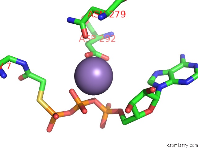
Mono view
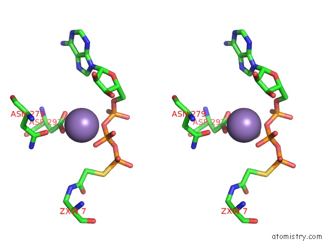
Stereo pair view
Mono view
Stereo pair view
A full contact list of Manganese with other atoms in the Mn binding
site number 1 of Crystal Structure of AKT1 (Aa 144-480) with A Bisubstrate within 5.0Å range:
|
Manganese binding site 2 out of 2 in 6buu
Go back to
Manganese binding site 2 out
of 2 in the Crystal Structure of AKT1 (Aa 144-480) with A Bisubstrate
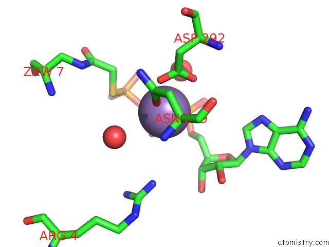
Mono view
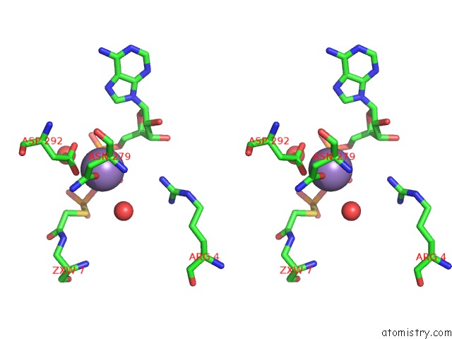
Stereo pair view
Mono view
Stereo pair view
A full contact list of Manganese with other atoms in the Mn binding
site number 2 of Crystal Structure of AKT1 (Aa 144-480) with A Bisubstrate within 5.0Å range:
|
Reference:
N.Chu,
A.L.Salguero,
A.Z.Liu,
Z.Chen,
D.R.Dempsey,
S.B.Ficarro,
W.M.Alexander,
J.A.Marto,
Y.Li,
L.M.Amzel,
S.B.Gabelli,
P.A.Cole.
Akt Kinase Activation Mechanisms Revealed Using Protein Semisynthesis. Cell V. 174 897 2018.
ISSN: ISSN 1097-4172
PubMed: 30078705
DOI: 10.1016/J.CELL.2018.07.003
Page generated: Sun Oct 6 03:57:02 2024
ISSN: ISSN 1097-4172
PubMed: 30078705
DOI: 10.1016/J.CELL.2018.07.003
Last articles
Zn in 9J0NZn in 9J0O
Zn in 9J0P
Zn in 9FJX
Zn in 9EKB
Zn in 9C0F
Zn in 9CAH
Zn in 9CH0
Zn in 9CH3
Zn in 9CH1