Manganese »
PDB 6a9v-6bh4 »
6b2u »
Manganese in PDB 6b2u: Crystal Structure of Xanthomonas Campestris Olea H285N with Cerulenin
Protein crystallography data
The structure of Crystal Structure of Xanthomonas Campestris Olea H285N with Cerulenin, PDB code: 6b2u
was solved by
M.R.Jensen,
B.R.Goblirsch,
M.A.Esler,
J.K.Christenson,
F.A.Mohamed,
L.P.Wackett,
C.M.Wilmot,
with X-Ray Crystallography technique. A brief refinement statistics is given in the table below:
| Resolution Low / High (Å) | 29.51 / 2.04 |
| Space group | P 32 2 1 |
| Cell size a, b, c (Å), α, β, γ (°) | 90.516, 90.516, 69.793, 90.00, 90.00, 120.00 |
| R / Rfree (%) | 17.9 / 23 |
Manganese Binding Sites:
The binding sites of Manganese atom in the Crystal Structure of Xanthomonas Campestris Olea H285N with Cerulenin
(pdb code 6b2u). This binding sites where shown within
5.0 Angstroms radius around Manganese atom.
In total only one binding site of Manganese was determined in the Crystal Structure of Xanthomonas Campestris Olea H285N with Cerulenin, PDB code: 6b2u:
In total only one binding site of Manganese was determined in the Crystal Structure of Xanthomonas Campestris Olea H285N with Cerulenin, PDB code: 6b2u:
Manganese binding site 1 out of 1 in 6b2u
Go back to
Manganese binding site 1 out
of 1 in the Crystal Structure of Xanthomonas Campestris Olea H285N with Cerulenin
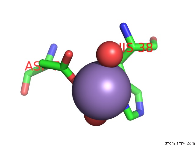
Mono view
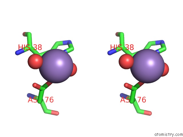
Stereo pair view
Mono view
Stereo pair view
A full contact list of Manganese with other atoms in the Mn binding
site number 1 of Crystal Structure of Xanthomonas Campestris Olea H285N with Cerulenin within 5.0Å range:
|
Reference:
M.R.Jensen,
B.R.Goblirsch,
M.A.Esler,
J.K.Christenson,
F.A.Mohamed,
L.P.Wackett,
C.M.Wilmot.
The Role of Olea HIS285 in Substrate Coordination of Long-Chain Acyl-Coa To Be Published.
Page generated: Sun Oct 6 03:54:00 2024
Last articles
Zn in 9J0NZn in 9J0O
Zn in 9J0P
Zn in 9FJX
Zn in 9EKB
Zn in 9C0F
Zn in 9CAH
Zn in 9CH0
Zn in 9CH3
Zn in 9CH1