Manganese »
PDB 6a9v-6bh4 »
6aob »
Manganese in PDB 6aob: Crystal Structure of Non-Canonical Dimeric Guanylyl Cyclase Domain of Rhogc Fusion Protein From the Aquatic Fungus Blastocladiella Emersonii
Protein crystallography data
The structure of Crystal Structure of Non-Canonical Dimeric Guanylyl Cyclase Domain of Rhogc Fusion Protein From the Aquatic Fungus Blastocladiella Emersonii, PDB code: 6aob
was solved by
R.Prem Kumar,
D.D.Oprian,
with X-Ray Crystallography technique. A brief refinement statistics is given in the table below:
| Resolution Low / High (Å) | 19.97 / 1.70 |
| Space group | C 2 2 21 |
| Cell size a, b, c (Å), α, β, γ (°) | 91.769, 95.880, 100.049, 90.00, 90.00, 90.00 |
| R / Rfree (%) | 16.4 / 19.6 |
Manganese Binding Sites:
The binding sites of Manganese atom in the Crystal Structure of Non-Canonical Dimeric Guanylyl Cyclase Domain of Rhogc Fusion Protein From the Aquatic Fungus Blastocladiella Emersonii
(pdb code 6aob). This binding sites where shown within
5.0 Angstroms radius around Manganese atom.
In total 2 binding sites of Manganese where determined in the Crystal Structure of Non-Canonical Dimeric Guanylyl Cyclase Domain of Rhogc Fusion Protein From the Aquatic Fungus Blastocladiella Emersonii, PDB code: 6aob:
Jump to Manganese binding site number: 1; 2;
In total 2 binding sites of Manganese where determined in the Crystal Structure of Non-Canonical Dimeric Guanylyl Cyclase Domain of Rhogc Fusion Protein From the Aquatic Fungus Blastocladiella Emersonii, PDB code: 6aob:
Jump to Manganese binding site number: 1; 2;
Manganese binding site 1 out of 2 in 6aob
Go back to
Manganese binding site 1 out
of 2 in the Crystal Structure of Non-Canonical Dimeric Guanylyl Cyclase Domain of Rhogc Fusion Protein From the Aquatic Fungus Blastocladiella Emersonii
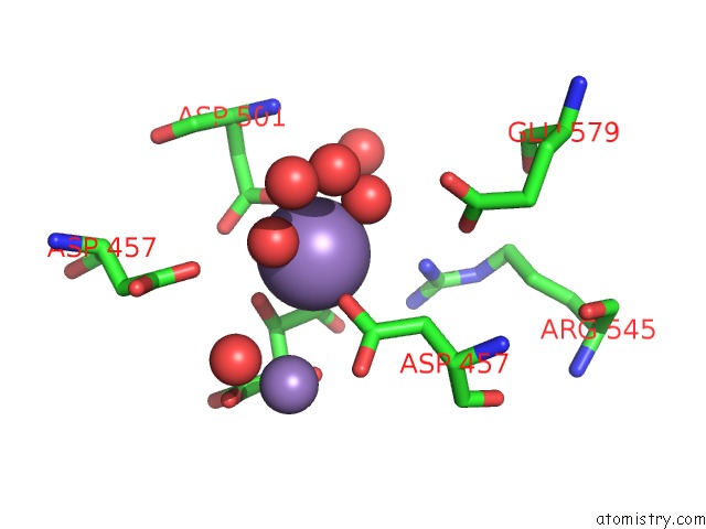
Mono view
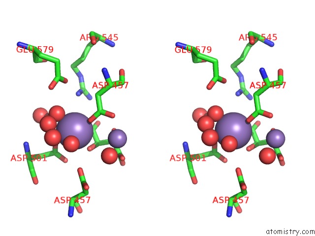
Stereo pair view
Mono view
Stereo pair view
A full contact list of Manganese with other atoms in the Mn binding
site number 1 of Crystal Structure of Non-Canonical Dimeric Guanylyl Cyclase Domain of Rhogc Fusion Protein From the Aquatic Fungus Blastocladiella Emersonii within 5.0Å range:
|
Manganese binding site 2 out of 2 in 6aob
Go back to
Manganese binding site 2 out
of 2 in the Crystal Structure of Non-Canonical Dimeric Guanylyl Cyclase Domain of Rhogc Fusion Protein From the Aquatic Fungus Blastocladiella Emersonii
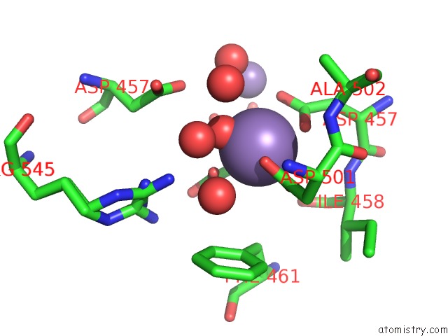
Mono view
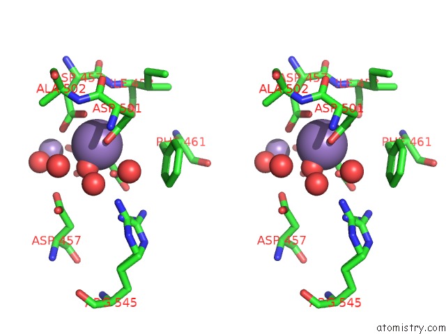
Stereo pair view
Mono view
Stereo pair view
A full contact list of Manganese with other atoms in the Mn binding
site number 2 of Crystal Structure of Non-Canonical Dimeric Guanylyl Cyclase Domain of Rhogc Fusion Protein From the Aquatic Fungus Blastocladiella Emersonii within 5.0Å range:
|
Reference:
R.P.Kumar,
B.R.Morehouse,
J.Fofana,
M.M.Trieu,
D.H.Zhou,
M.O.Lorenz,
D.D.Oprian.
Structure and Monomer/Dimer Equilibrium For the Guanylyl Cyclase Domain of the Optogenetics Protein Rhogc. J. Biol. Chem. V. 292 21578 2017.
ISSN: ESSN 1083-351X
PubMed: 29118188
DOI: 10.1074/JBC.M117.812685
Page generated: Sun Oct 6 03:51:26 2024
ISSN: ESSN 1083-351X
PubMed: 29118188
DOI: 10.1074/JBC.M117.812685
Last articles
Zn in 9J0NZn in 9J0O
Zn in 9J0P
Zn in 9FJX
Zn in 9EKB
Zn in 9C0F
Zn in 9CAH
Zn in 9CH0
Zn in 9CH3
Zn in 9CH1