Manganese »
PDB 6a9v-6bh4 »
6adx »
Manganese in PDB 6adx: Crystal Structure of the Zika Virus NS3 Helicase (Adp-MN2+ Complex, Form 1)
Enzymatic activity of Crystal Structure of the Zika Virus NS3 Helicase (Adp-MN2+ Complex, Form 1)
All present enzymatic activity of Crystal Structure of the Zika Virus NS3 Helicase (Adp-MN2+ Complex, Form 1):
3.4.21.91; 3.6.1.15; 3.6.4.13;
3.4.21.91; 3.6.1.15; 3.6.4.13;
Protein crystallography data
The structure of Crystal Structure of the Zika Virus NS3 Helicase (Adp-MN2+ Complex, Form 1), PDB code: 6adx
was solved by
J.Fang,
G.Lu,
P.Gong,
with X-Ray Crystallography technique. A brief refinement statistics is given in the table below:
| Resolution Low / High (Å) | 29.39 / 1.75 |
| Space group | P 1 21 1 |
| Cell size a, b, c (Å), α, β, γ (°) | 53.750, 68.658, 56.929, 90.00, 92.50, 90.00 |
| R / Rfree (%) | 17.9 / 21.1 |
Manganese Binding Sites:
The binding sites of Manganese atom in the Crystal Structure of the Zika Virus NS3 Helicase (Adp-MN2+ Complex, Form 1)
(pdb code 6adx). This binding sites where shown within
5.0 Angstroms radius around Manganese atom.
In total only one binding site of Manganese was determined in the Crystal Structure of the Zika Virus NS3 Helicase (Adp-MN2+ Complex, Form 1), PDB code: 6adx:
In total only one binding site of Manganese was determined in the Crystal Structure of the Zika Virus NS3 Helicase (Adp-MN2+ Complex, Form 1), PDB code: 6adx:
Manganese binding site 1 out of 1 in 6adx
Go back to
Manganese binding site 1 out
of 1 in the Crystal Structure of the Zika Virus NS3 Helicase (Adp-MN2+ Complex, Form 1)
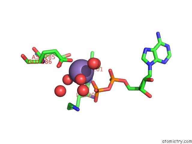
Mono view
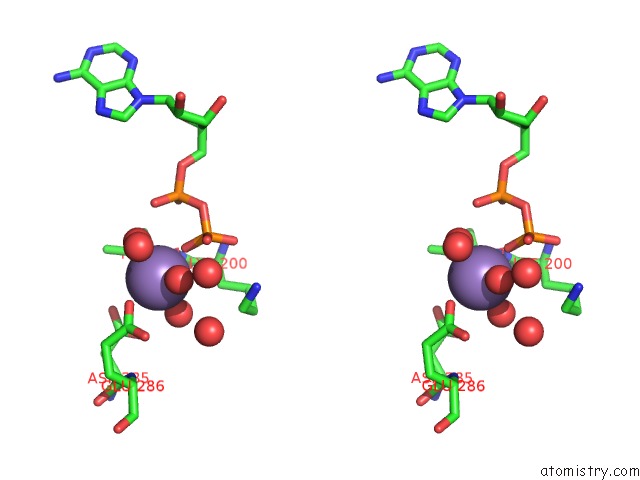
Stereo pair view
Mono view
Stereo pair view
A full contact list of Manganese with other atoms in the Mn binding
site number 1 of Crystal Structure of the Zika Virus NS3 Helicase (Adp-MN2+ Complex, Form 1) within 5.0Å range:
|
Reference:
J.Fang,
X.Jing,
G.Lu,
Y.Xu,
P.Gong.
Crystallographic Snapshots of the Zika Virus NS3 Helicase Help Visualize the Reactant Water Replenishment. Acs Infect Dis V. 5 177 2019.
ISSN: ESSN 2373-8227
PubMed: 30672289
DOI: 10.1021/ACSINFECDIS.8B00214
Page generated: Sun Oct 6 03:49:52 2024
ISSN: ESSN 2373-8227
PubMed: 30672289
DOI: 10.1021/ACSINFECDIS.8B00214
Last articles
Zn in 9J0NZn in 9J0O
Zn in 9J0P
Zn in 9FJX
Zn in 9EKB
Zn in 9C0F
Zn in 9CAH
Zn in 9CH0
Zn in 9CH3
Zn in 9CH1