Manganese »
PDB 6a9v-6bh4 »
6a9v »
Manganese in PDB 6a9v: Crystal Structure of ICP55 From Saccharomyces Cerevisiae (N-Terminal 42 Residues Deletion)
Enzymatic activity of Crystal Structure of ICP55 From Saccharomyces Cerevisiae (N-Terminal 42 Residues Deletion)
All present enzymatic activity of Crystal Structure of ICP55 From Saccharomyces Cerevisiae (N-Terminal 42 Residues Deletion):
3.4.11.26;
3.4.11.26;
Protein crystallography data
The structure of Crystal Structure of ICP55 From Saccharomyces Cerevisiae (N-Terminal 42 Residues Deletion), PDB code: 6a9v
was solved by
R.Singh,
A.Kumar,
V.D.Goyal,
R.D.Makde,
with X-Ray Crystallography technique. A brief refinement statistics is given in the table below:
| Resolution Low / High (Å) | 45.55 / 2.90 |
| Space group | I 4 2 2 |
| Cell size a, b, c (Å), α, β, γ (°) | 142.081, 142.081, 118.731, 90.00, 90.00, 90.00 |
| R / Rfree (%) | 21.3 / 22.5 |
Manganese Binding Sites:
The binding sites of Manganese atom in the Crystal Structure of ICP55 From Saccharomyces Cerevisiae (N-Terminal 42 Residues Deletion)
(pdb code 6a9v). This binding sites where shown within
5.0 Angstroms radius around Manganese atom.
In total 2 binding sites of Manganese where determined in the Crystal Structure of ICP55 From Saccharomyces Cerevisiae (N-Terminal 42 Residues Deletion), PDB code: 6a9v:
Jump to Manganese binding site number: 1; 2;
In total 2 binding sites of Manganese where determined in the Crystal Structure of ICP55 From Saccharomyces Cerevisiae (N-Terminal 42 Residues Deletion), PDB code: 6a9v:
Jump to Manganese binding site number: 1; 2;
Manganese binding site 1 out of 2 in 6a9v
Go back to
Manganese binding site 1 out
of 2 in the Crystal Structure of ICP55 From Saccharomyces Cerevisiae (N-Terminal 42 Residues Deletion)
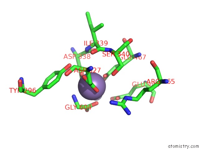
Mono view
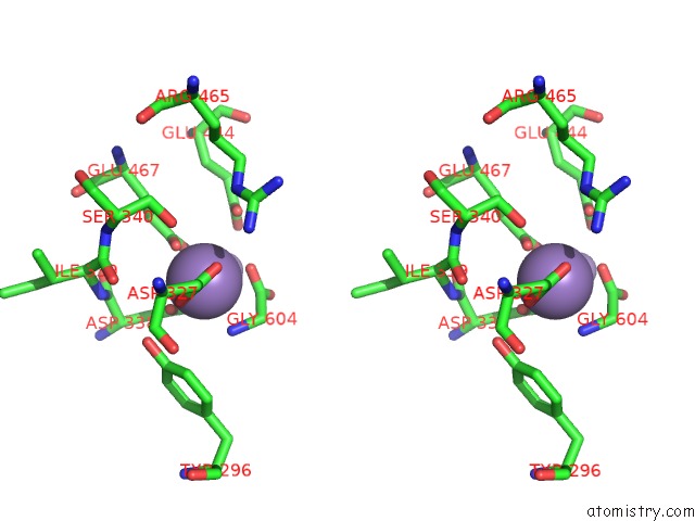
Stereo pair view
Mono view
Stereo pair view
A full contact list of Manganese with other atoms in the Mn binding
site number 1 of Crystal Structure of ICP55 From Saccharomyces Cerevisiae (N-Terminal 42 Residues Deletion) within 5.0Å range:
|
Manganese binding site 2 out of 2 in 6a9v
Go back to
Manganese binding site 2 out
of 2 in the Crystal Structure of ICP55 From Saccharomyces Cerevisiae (N-Terminal 42 Residues Deletion)
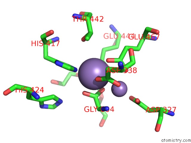
Mono view
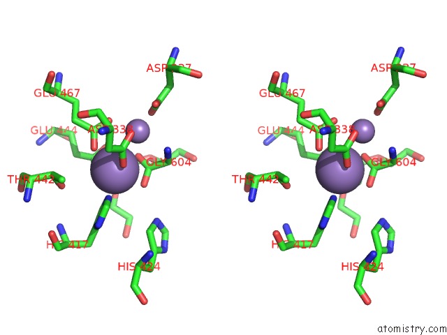
Stereo pair view
Mono view
Stereo pair view
A full contact list of Manganese with other atoms in the Mn binding
site number 2 of Crystal Structure of ICP55 From Saccharomyces Cerevisiae (N-Terminal 42 Residues Deletion) within 5.0Å range:
|
Reference:
R.Singh,
V.D.Goyal,
A.Kumar,
N.S.Sabharwal,
R.D.Makde.
Crystal Structures and Biochemical Analyses of Intermediate Cleavage Peptidase: Role of Dynamics in Enzymatic Function. Febs Lett. V. 593 443 2019.
ISSN: ISSN 1873-3468
PubMed: 30582634
DOI: 10.1002/1873-3468.13321
Page generated: Sun Oct 6 03:49:52 2024
ISSN: ISSN 1873-3468
PubMed: 30582634
DOI: 10.1002/1873-3468.13321
Last articles
Zn in 9J0NZn in 9J0O
Zn in 9J0P
Zn in 9FJX
Zn in 9EKB
Zn in 9C0F
Zn in 9CAH
Zn in 9CH0
Zn in 9CH3
Zn in 9CH1