Manganese »
PDB 5z2k-6a9u »
5zye »
Manganese in PDB 5zye: Crystal Structure of Glucose Isomerase Soaked with MN2+ and Glucose
Enzymatic activity of Crystal Structure of Glucose Isomerase Soaked with MN2+ and Glucose
All present enzymatic activity of Crystal Structure of Glucose Isomerase Soaked with MN2+ and Glucose:
5.3.1.5;
5.3.1.5;
Protein crystallography data
The structure of Crystal Structure of Glucose Isomerase Soaked with MN2+ and Glucose, PDB code: 5zye
was solved by
K.H.Nam,
with X-Ray Crystallography technique. A brief refinement statistics is given in the table below:
| Resolution Low / High (Å) | 71.19 / 1.40 |
| Space group | I 2 2 2 |
| Cell size a, b, c (Å), α, β, γ (°) | 92.749, 98.961, 102.481, 90.00, 90.00, 90.00 |
| R / Rfree (%) | 16.7 / 18.6 |
Manganese Binding Sites:
The binding sites of Manganese atom in the Crystal Structure of Glucose Isomerase Soaked with MN2+ and Glucose
(pdb code 5zye). This binding sites where shown within
5.0 Angstroms radius around Manganese atom.
In total 2 binding sites of Manganese where determined in the Crystal Structure of Glucose Isomerase Soaked with MN2+ and Glucose, PDB code: 5zye:
Jump to Manganese binding site number: 1; 2;
In total 2 binding sites of Manganese where determined in the Crystal Structure of Glucose Isomerase Soaked with MN2+ and Glucose, PDB code: 5zye:
Jump to Manganese binding site number: 1; 2;
Manganese binding site 1 out of 2 in 5zye
Go back to
Manganese binding site 1 out
of 2 in the Crystal Structure of Glucose Isomerase Soaked with MN2+ and Glucose
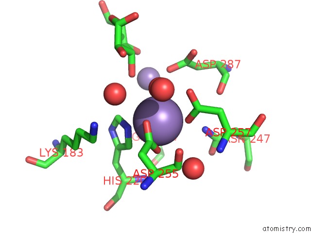
Mono view
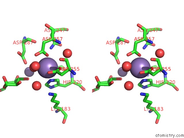
Stereo pair view
Mono view
Stereo pair view
A full contact list of Manganese with other atoms in the Mn binding
site number 1 of Crystal Structure of Glucose Isomerase Soaked with MN2+ and Glucose within 5.0Å range:
|
Manganese binding site 2 out of 2 in 5zye
Go back to
Manganese binding site 2 out
of 2 in the Crystal Structure of Glucose Isomerase Soaked with MN2+ and Glucose
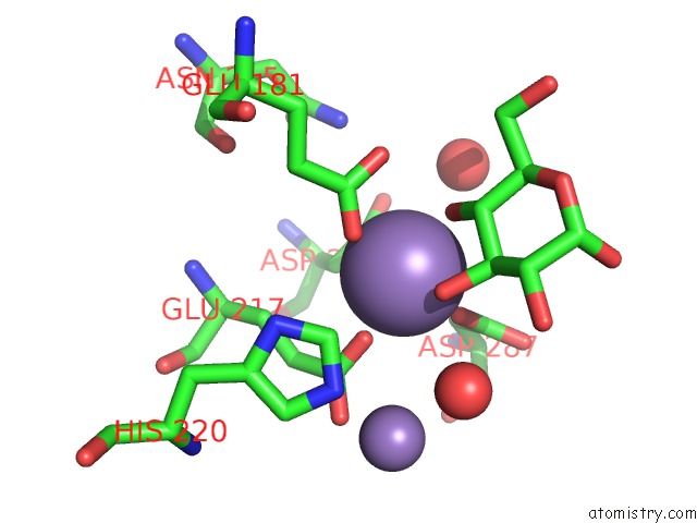
Mono view
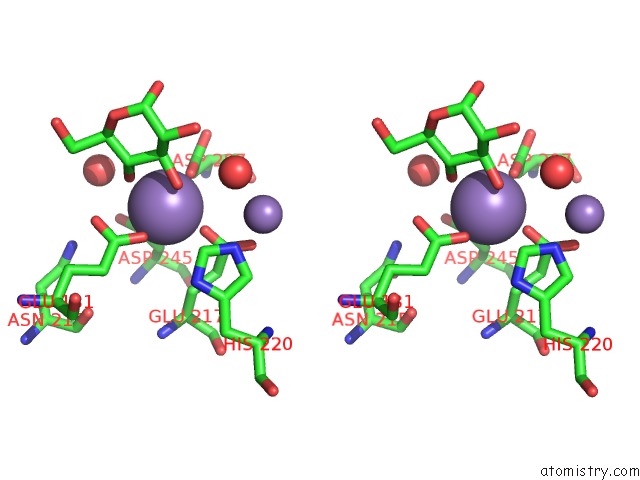
Stereo pair view
Mono view
Stereo pair view
A full contact list of Manganese with other atoms in the Mn binding
site number 2 of Crystal Structure of Glucose Isomerase Soaked with MN2+ and Glucose within 5.0Å range:
|
Reference:
J.E.Bae,
K.Y.Hwang,
K.H.Nam.
Structural Analysis of Substrate Recognition By Glucose Isomerase in MN2+Binding Mode at M2 Site in S. Rubiginosus Biochem. Biophys. Res. V. 503 770 2018COMMUN..
ISSN: ESSN 1090-2104
PubMed: 29909012
DOI: 10.1016/J.BBRC.2018.06.074
Page generated: Sun Oct 6 03:45:34 2024
ISSN: ESSN 1090-2104
PubMed: 29909012
DOI: 10.1016/J.BBRC.2018.06.074
Last articles
Zn in 9J0NZn in 9J0O
Zn in 9J0P
Zn in 9FJX
Zn in 9EKB
Zn in 9C0F
Zn in 9CAH
Zn in 9CH0
Zn in 9CH3
Zn in 9CH1