Atomistry » Manganese » PDB 5z2k-6a9u » 5zt0
Atomistry »
  Manganese »
    PDB 5z2k-6a9u »
      5zt0 »

Manganese in PDB 5zt0: Crystal Structure of Protein Phosphate 1 Complexed with PP1 Binding Domain of Gl

Enzymatic activity of Crystal Structure of Protein Phosphate 1 Complexed with PP1 Binding Domain of Gl

All present enzymatic activity of Crystal Structure of Protein Phosphate 1 Complexed with PP1 Binding Domain of Gl:
3.1.3.16;

Protein crystallography data

The structure of Crystal Structure of Protein Phosphate 1 Complexed with PP1 Binding Domain of Gl, PDB code: 5zt0 was solved by J.Yu, S.Xiang, with X-Ray Crystallography technique. A brief refinement statistics is given in the table below:

Resolution Low / High (Å) 41.79 / 3.32
Space group P 32
Cell size a, b, c (Å), α, β, γ (°) 106.530, 106.530, 187.510, 90.00, 90.00, 120.00
R / Rfree (%) 20 / 23.3

Manganese Binding Sites:

Pages:

>>> Page 1 <<< Page 2, Binding sites: 11 - 12;

Binding sites:

The binding sites of Manganese atom in the Crystal Structure of Protein Phosphate 1 Complexed with PP1 Binding Domain of Gl (pdb code 5zt0). This binding sites where shown within 5.0 Angstroms radius around Manganese atom.
In total 12 binding sites of Manganese where determined in the Crystal Structure of Protein Phosphate 1 Complexed with PP1 Binding Domain of Gl, PDB code: 5zt0:
Jump to Manganese binding site number: 1; 2; 3; 4; 5; 6; 7; 8; 9; 10;

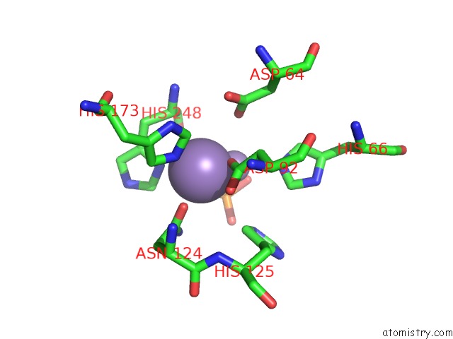
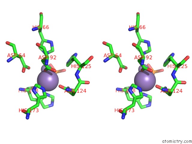
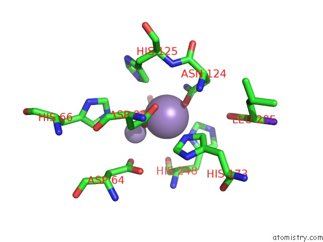
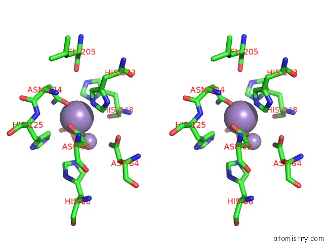
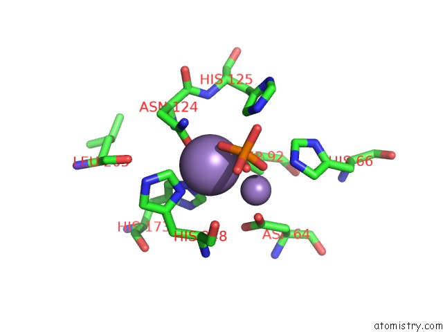
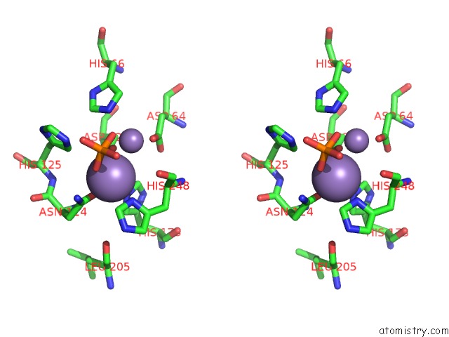
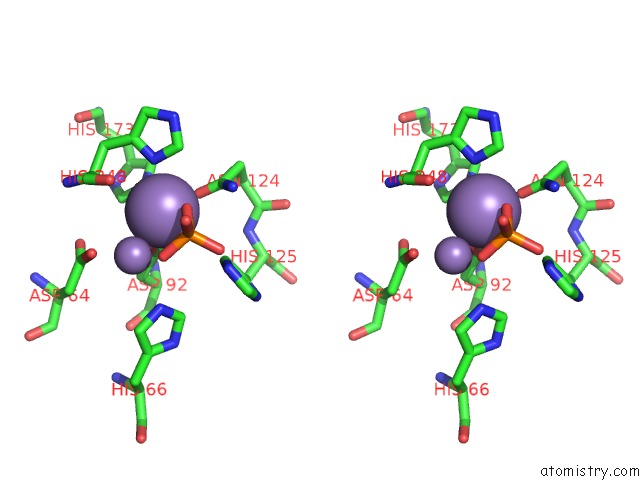
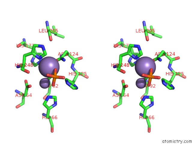
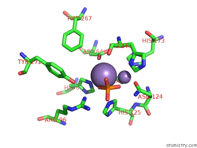
Manganese binding site 1 out of 12 in 5zt0

Go back to Manganese Binding Sites List in 5zt0
Manganese binding site 1 out of 12 in the Crystal Structure of Protein Phosphate 1 Complexed with PP1 Binding Domain of Gl


Mono view


Stereo pair view

A full contact list of Manganese with other atoms in the Mn binding site number 1 of Crystal Structure of Protein Phosphate 1 Complexed with PP1 Binding Domain of Gl within 5.0Å range:
probe atom residue distance (Å) B Occ
A:Mn401

b:47.4
occ:1.00
OD2 A:ASP92 1.9 0.6 1.0
MN A:MN402 2.0 31.8 0.5
NE2 A:HIS173 2.5 90.1 1.0
ND1 A:HIS248 2.5 91.6 1.0
OD1 A:ASN124 2.5 76.1 1.0
O3 A:PO4403 2.7 29.6 0.8
CG A:ASP92 2.8 71.8 1.0
CE1 A:HIS173 3.1 85.1 1.0
OD1 A:ASP92 3.1 71.7 1.0
CE1 A:HIS248 3.2 82.7 1.0
CG A:ASN124 3.3 76.6 1.0
ND2 A:ASN124 3.5 76.8 1.0
CA A:HIS248 3.5 74.5 1.0
CG A:HIS248 3.6 88.2 1.0
CD2 A:HIS173 3.6 97.3 1.0
OD2 A:ASP64 3.6 74.0 1.0
O A:HIS248 3.8 74.6 1.0
P A:PO4403 3.8 52.0 0.8
CD2 A:HIS125 4.0 74.8 1.0
O4 A:PO4403 4.0 58.4 0.8
CB A:HIS248 4.0 75.5 1.0
C A:HIS248 4.1 75.0 1.0
CB A:ASP92 4.2 0.8 1.0
NE2 A:HIS66 4.3 72.1 1.0
NE2 A:HIS125 4.4 75.0 1.0
ND1 A:HIS173 4.4 91.3 1.0
CG A:ASP64 4.4 73.3 1.0
NE2 A:HIS248 4.4 89.1 1.0
O2 A:PO4403 4.4 66.7 0.8
OD1 A:ASP64 4.4 95.8 1.0
N A:HIS248 4.6 74.9 1.0
CD2 A:HIS248 4.6 76.2 1.0
CG A:HIS173 4.6 96.1 1.0
CB A:ASN124 4.8 77.3 1.0
CE1 A:HIS66 4.8 71.2 1.0
N A:ASN124 4.9 76.4 1.0

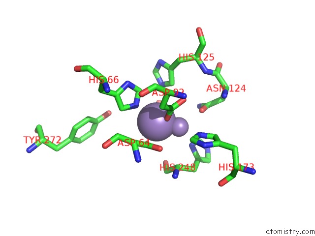
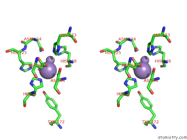
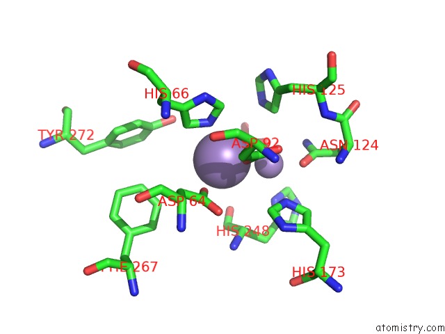
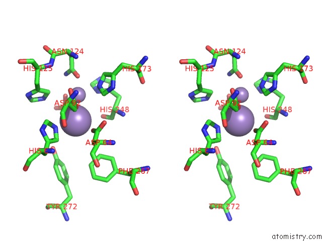
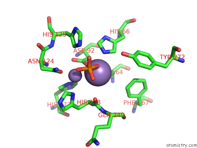
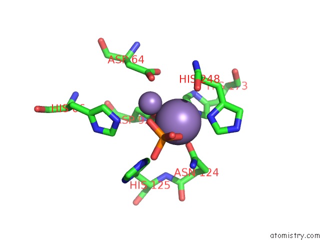
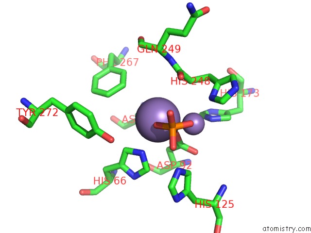
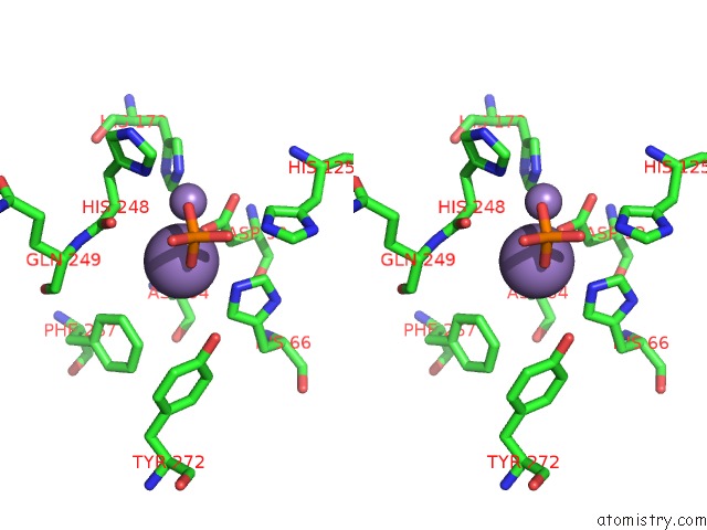
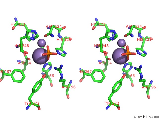
Manganese binding site 2 out of 12 in 5zt0

Go back to Manganese Binding Sites List in 5zt0
Manganese binding site 2 out of 12 in the Crystal Structure of Protein Phosphate 1 Complexed with PP1 Binding Domain of Gl


Mono view


Stereo pair view

A full contact list of Manganese with other atoms in the Mn binding site number 2 of Crystal Structure of Protein Phosphate 1 Complexed with PP1 Binding Domain of Gl within 5.0Å range:
probe atom residue distance (Å) B Occ
A:Mn402

b:31.8
occ:0.50
OD2 A:ASP92 1.8 0.6 1.0
MN A:MN401 2.0 47.4 1.0
O4 A:PO4403 2.3 58.4 0.8
O3 A:PO4403 2.5 29.6 0.8
NE2 A:HIS66 2.6 72.1 1.0
P A:PO4403 2.9 52.0 0.8
OD2 A:ASP64 2.9 74.0 1.0
CG A:ASP92 3.0 71.8 1.0
CE1 A:HIS66 3.4 71.2 1.0
NE2 A:HIS125 3.6 75.0 1.0
O2 A:PO4403 3.7 66.7 0.8
CD2 A:HIS125 3.7 74.8 1.0
CD2 A:HIS66 3.7 71.9 1.0
OD1 A:ASP92 3.7 71.7 1.0
CG A:ASP64 3.9 73.3 1.0
CB A:ASP92 3.9 0.8 1.0
ND1 A:HIS248 4.0 91.6 1.0
O A:HIS248 4.1 74.6 1.0
OD1 A:ASN124 4.1 76.1 1.0
O1 A:PO4403 4.2 34.5 0.8
NE2 A:HIS173 4.3 90.1 1.0
CA A:HIS248 4.4 74.5 1.0
C A:HIS248 4.5 75.0 1.0
OD1 A:ASP64 4.5 95.8 1.0
CE1 A:HIS173 4.5 85.1 1.0
ND1 A:HIS66 4.6 70.5 1.0
OH A:TYR272 4.6 0.5 1.0
ND2 A:ASN124 4.6 76.8 1.0
CE1 A:HIS248 4.6 82.7 1.0
CE1 A:HIS125 4.8 74.9 1.0
CG A:HIS66 4.8 70.8 1.0
CG A:ASN124 4.8 76.6 1.0
CB A:ASP64 4.9 72.7 1.0
CG A:HIS125 4.9 74.5 1.0

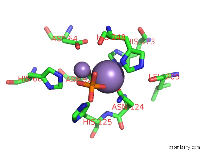
Manganese binding site 3 out of 12 in 5zt0

Go back to Manganese Binding Sites List in 5zt0
Manganese binding site 3 out of 12 in the Crystal Structure of Protein Phosphate 1 Complexed with PP1 Binding Domain of Gl


Mono view


Stereo pair view

A full contact list of Manganese with other atoms in the Mn binding site number 3 of Crystal Structure of Protein Phosphate 1 Complexed with PP1 Binding Domain of Gl within 5.0Å range:
probe atom residue distance (Å) B Occ
B:Mn401

b:0.6
occ:1.00
OD1 B:ASN124 2.1 80.4 1.0
OD2 B:ASP92 2.2 84.8 1.0
O3 B:PO4403 2.3 18.6 0.8
ND1 B:HIS248 2.7 0.2 1.0
MN B:MN402 2.8 54.6 0.5
CG B:ASN124 2.8 81.1 1.0
ND2 B:ASN124 2.9 81.8 1.0
CG B:ASP92 2.9 82.8 1.0
NE2 B:HIS173 2.9 0.6 1.0
OD1 B:ASP92 3.0 82.6 1.0
CE1 B:HIS248 3.1 0.4 1.0
P B:PO4403 3.2 68.4 0.8
CD2 B:HIS125 3.3 0.4 1.0
O2 B:PO4403 3.4 71.6 0.8
O4 B:PO4403 3.6 83.5 0.8
CE1 B:HIS173 3.7 0.6 1.0
NE2 B:HIS125 3.8 81.2 1.0
CG B:HIS248 4.0 0.8 1.0
CD2 B:HIS173 4.0 97.5 1.0
O B:HIS248 4.3 91.3 1.0
CA B:HIS248 4.3 97.0 1.0
CB B:ASN124 4.3 81.1 1.0
NE2 B:HIS248 4.4 0.6 1.0
CB B:ASP92 4.4 85.5 1.0
OD2 B:ASP64 4.5 97.2 1.0
NE2 B:HIS66 4.6 70.9 1.0
CG B:HIS125 4.6 79.8 1.0
O1 B:PO4403 4.6 61.2 0.8
CB B:HIS248 4.6 0.8 1.0
N B:ASN124 4.7 79.3 1.0
C B:HIS248 4.8 96.2 1.0
CD2 B:HIS248 4.8 0.6 1.0
N B:HIS125 4.9 79.2 1.0
O B:LEU205 4.9 92.9 1.0
ND1 B:HIS173 4.9 0.6 1.0
CE1 B:HIS66 5.0 70.5 1.0

Manganese binding site 4 out of 12 in 5zt0

Go back to Manganese Binding Sites List in 5zt0
Manganese binding site 4 out of 12 in the Crystal Structure of Protein Phosphate 1 Complexed with PP1 Binding Domain of Gl


Mono view


Stereo pair view

A full contact list of Manganese with other atoms in the Mn binding site number 4 of Crystal Structure of Protein Phosphate 1 Complexed with PP1 Binding Domain of Gl within 5.0Å range:
probe atom residue distance (Å) B Occ
B:Mn402

b:54.6
occ:0.50
OD2 B:ASP92 2.1 84.8 1.0
O2 B:PO4403 2.2 71.6 0.8
OD2 B:ASP64 2.6 97.2 1.0
NE2 B:HIS66 2.7 70.9 1.0
MN B:MN401 2.8 0.6 1.0
P B:PO4403 3.3 68.4 0.8
CG B:ASP92 3.3 82.8 1.0
CD2 B:HIS66 3.4 70.5 1.0
O3 B:PO4403 3.6 18.6 0.8
CG B:ASP64 3.6 91.9 1.0
O4 B:PO4403 3.7 83.5 0.8
CE1 B:HIS66 3.8 70.5 1.0
O B:HIS248 3.9 91.3 1.0
CB B:ASP92 4.1 85.5 1.0
OH B:TYR272 4.1 0.1 1.0
OD1 B:ASP92 4.2 82.6 1.0
NE2 B:HIS125 4.3 81.2 1.0
OD1 B:ASP64 4.3 95.2 1.0
CD2 B:HIS125 4.3 0.4 1.0
C B:HIS248 4.3 96.2 1.0
ND1 B:HIS248 4.4 0.2 1.0
CA B:HIS248 4.4 97.0 1.0
CB B:ASP64 4.6 74.4 1.0
O1 B:PO4403 4.6 61.2 0.8
CE1 B:PHE267 4.7 0.7 1.0
CG B:HIS66 4.7 69.8 1.0
NE2 B:HIS173 4.7 0.6 1.0
CE1 B:HIS173 4.8 0.6 1.0
ND1 B:HIS66 4.8 69.8 1.0
OD1 B:ASN124 4.8 80.4 1.0
CZ B:TYR272 4.9 0.8 1.0

Manganese binding site 5 out of 12 in 5zt0

Go back to Manganese Binding Sites List in 5zt0
Manganese binding site 5 out of 12 in the Crystal Structure of Protein Phosphate 1 Complexed with PP1 Binding Domain of Gl


Mono view


Stereo pair view

A full contact list of Manganese with other atoms in the Mn binding site number 5 of Crystal Structure of Protein Phosphate 1 Complexed with PP1 Binding Domain of Gl within 5.0Å range:
probe atom residue distance (Å) B Occ
C:Mn401

b:65.7
occ:1.00
OD1 C:ASN124 2.0 0.2 1.0
O3 C:PO4403 2.1 18.4 0.8
OD2 C:ASP92 2.4 74.1 1.0
ND1 C:HIS248 2.4 99.8 1.0
NE2 C:HIS173 2.7 68.5 1.0
CG C:ASN124 2.7 71.0 1.0
ND2 C:ASN124 2.9 71.5 1.0
CE1 C:HIS248 2.9 96.1 1.0
MN C:MN402 2.9 21.6 0.5
CG C:ASP92 3.1 74.0 1.0
OD1 C:ASP92 3.1 74.4 1.0
P C:PO4403 3.2 65.0 0.8
O1 C:PO4403 3.3 67.5 0.8
CE1 C:HIS173 3.5 68.9 1.0
CG C:HIS248 3.6 76.3 1.0
CD2 C:HIS125 3.7 81.1 1.0
CD2 C:HIS173 3.8 69.7 1.0
O C:HIS248 4.0 97.2 1.0
O4 C:PO4403 4.0 77.3 0.8
CA C:HIS248 4.0 75.3 1.0
OD2 C:ASP64 4.1 73.5 1.0
NE2 C:HIS248 4.1 86.6 1.0
NE2 C:HIS125 4.1 70.9 1.0
CB C:ASN124 4.2 71.1 1.0
CB C:HIS248 4.3 76.4 1.0
O2 C:PO4403 4.4 0.5 0.8
C C:HIS248 4.5 75.6 1.0
CB C:ASP92 4.5 96.6 1.0
CD2 C:HIS248 4.5 88.0 1.0
N C:ASN124 4.6 70.0 1.0
ND1 C:HIS173 4.7 70.4 1.0
NE2 C:HIS66 4.8 85.4 1.0
O C:LEU205 4.8 71.7 1.0
CA C:ASN124 4.9 70.5 1.0
CG C:HIS173 4.9 71.2 1.0
CG C:HIS125 4.9 72.2 1.0
CG C:ASP64 4.9 73.2 1.0

Manganese binding site 6 out of 12 in 5zt0

Go back to Manganese Binding Sites List in 5zt0
Manganese binding site 6 out of 12 in the Crystal Structure of Protein Phosphate 1 Complexed with PP1 Binding Domain of Gl


Mono view


Stereo pair view

A full contact list of Manganese with other atoms in the Mn binding site number 6 of Crystal Structure of Protein Phosphate 1 Complexed with PP1 Binding Domain of Gl within 5.0Å range:
probe atom residue distance (Å) B Occ
C:Mn402

b:21.6
occ:0.50
OD2 C:ASP92 1.9 74.1 1.0
OD2 C:ASP64 1.9 73.5 1.0
O1 C:PO4403 2.1 67.5 0.8
NE2 C:HIS66 2.7 85.4 1.0
MN C:MN401 2.9 65.7 1.0
CG C:ASP64 3.0 73.2 1.0
CG C:ASP92 3.0 74.0 1.0
P C:PO4403 3.2 65.0 0.8
O3 C:PO4403 3.5 18.4 0.8
CD2 C:HIS66 3.5 73.7 1.0
O C:HIS248 3.6 97.2 1.0
CB C:ASP92 3.7 96.6 1.0
CE1 C:HIS66 3.7 74.1 1.0
OD1 C:ASP64 3.8 74.8 1.0
O4 C:PO4403 3.9 77.3 0.8
CB C:ASP64 3.9 88.0 1.0
C C:HIS248 4.0 75.6 1.0
CA C:HIS248 4.0 75.3 1.0
OD1 C:ASP92 4.0 74.4 1.0
ND1 C:HIS248 4.3 99.8 1.0
CE1 C:HIS173 4.3 68.9 1.0
CE1 C:PHE267 4.4 82.7 1.0
NE2 C:HIS173 4.4 68.5 1.0
O2 C:PO4403 4.4 0.5 0.8
CD2 C:HIS125 4.5 81.1 1.0
NE2 C:HIS125 4.6 70.9 1.0
OH C:TYR272 4.6 84.3 1.0
CG C:HIS66 4.7 73.6 1.0
N C:HIS248 4.7 79.2 1.0
OD1 C:ASN124 4.7 0.2 1.0
ND1 C:HIS66 4.8 73.9 1.0
CZ C:PHE267 4.8 83.3 1.0
N C:GLN249 4.9 76.5 1.0

Manganese binding site 7 out of 12 in 5zt0

Go back to Manganese Binding Sites List in 5zt0
Manganese binding site 7 out of 12 in the Crystal Structure of Protein Phosphate 1 Complexed with PP1 Binding Domain of Gl


Mono view


Stereo pair view

A full contact list of Manganese with other atoms in the Mn binding site number 7 of Crystal Structure of Protein Phosphate 1 Complexed with PP1 Binding Domain of Gl within 5.0Å range:
probe atom residue distance (Å) B Occ
D:Mn401

b:47.3
occ:1.00
OD2 D:ASP92 2.0 77.3 1.0
O1 D:PO4403 2.3 25.9 0.8
OD1 D:ASN124 2.4 78.0 1.0
ND1 D:HIS248 2.5 91.4 1.0
NE2 D:HIS173 2.6 0.6 1.0
MN D:MN402 2.7 56.4 0.5
CG D:ASP92 2.8 75.9 1.0
OD1 D:ASP92 3.0 75.2 1.0
CG D:ASN124 3.3 78.7 1.0
CE1 D:HIS173 3.3 0.1 1.0
CE1 D:HIS248 3.3 85.6 1.0
P D:PO4403 3.4 65.1 0.8
O3 D:PO4403 3.4 49.1 0.8
ND2 D:ASN124 3.4 79.2 1.0
CA D:HIS248 3.6 81.3 1.0
CG D:HIS248 3.6 82.2 1.0
O D:HIS248 3.8 82.5 1.0
CD2 D:HIS173 3.8 0.7 1.0
OD2 D:ASP64 3.8 83.4 1.0
CD2 D:HIS125 3.8 84.8 1.0
CB D:HIS248 4.0 82.2 1.0
NE2 D:HIS125 4.1 78.0 1.0
C D:HIS248 4.1 85.0 1.0
O2 D:PO4403 4.2 0.5 0.8
CB D:ASP92 4.3 83.5 1.0
NE2 D:HIS248 4.5 83.2 1.0
NE2 D:HIS66 4.5 88.6 1.0
CG D:ASP64 4.5 93.3 1.0
ND1 D:HIS173 4.5 0.7 1.0
O4 D:PO4403 4.6 65.3 0.8
N D:HIS248 4.6 81.3 1.0
OD1 D:ASP64 4.6 0.2 1.0
CD2 D:HIS248 4.7 85.2 1.0
CB D:ASN124 4.7 79.2 1.0
CG D:HIS173 4.8 0.0 1.0
N D:ASN124 5.0 0.0 1.0

Manganese binding site 8 out of 12 in 5zt0

Go back to Manganese Binding Sites List in 5zt0
Manganese binding site 8 out of 12 in the Crystal Structure of Protein Phosphate 1 Complexed with PP1 Binding Domain of Gl


Mono view


Stereo pair view

A full contact list of Manganese with other atoms in the Mn binding site number 8 of Crystal Structure of Protein Phosphate 1 Complexed with PP1 Binding Domain of Gl within 5.0Å range:
probe atom residue distance (Å) B Occ
D:Mn402

b:56.4
occ:0.50
O3 D:PO4403 2.0 49.1 0.8
OD2 D:ASP64 2.2 83.4 1.0
OD2 D:ASP92 2.2 77.3 1.0
MN D:MN401 2.7 47.3 1.0
NE2 D:HIS66 2.8 88.6 1.0
P D:PO4403 3.2 65.1 0.8
CG D:ASP64 3.3 93.3 1.0
O1 D:PO4403 3.3 25.9 0.8
CG D:ASP92 3.4 75.9 1.0
CD2 D:HIS66 3.5 93.3 1.0
O D:HIS248 3.8 82.5 1.0
CE1 D:HIS66 3.9 85.7 1.0
C D:HIS248 4.0 85.0 1.0
OD1 D:ASP64 4.1 0.2 1.0
CB D:ASP92 4.1 83.5 1.0
CE1 D:PHE267 4.1 0.9 1.0
CB D:ASP64 4.1 94.0 1.0
CA D:HIS248 4.1 81.3 1.0
O2 D:PO4403 4.2 0.5 0.8
O4 D:PO4403 4.2 65.3 0.8
OH D:TYR272 4.3 0.5 1.0
OD1 D:ASP92 4.4 75.2 1.0
NE2 D:HIS125 4.5 78.0 1.0
ND1 D:HIS248 4.5 91.4 1.0
CZ D:PHE267 4.6 0.6 1.0
CD2 D:HIS125 4.7 84.8 1.0
CE1 D:HIS173 4.7 0.1 1.0
NE2 D:HIS173 4.7 0.6 1.0
CG D:HIS66 4.7 0.7 1.0
N D:HIS248 4.8 81.3 1.0
N D:GLN249 4.8 88.0 1.0
ND1 D:HIS66 4.9 99.6 1.0
CZ D:TYR272 4.9 0.4 1.0

Manganese binding site 9 out of 12 in 5zt0

Go back to Manganese Binding Sites List in 5zt0
Manganese binding site 9 out of 12 in the Crystal Structure of Protein Phosphate 1 Complexed with PP1 Binding Domain of Gl


Mono view


Stereo pair view

A full contact list of Manganese with other atoms in the Mn binding site number 9 of Crystal Structure of Protein Phosphate 1 Complexed with PP1 Binding Domain of Gl within 5.0Å range:
probe atom residue distance (Å) B Occ
E:Mn401

b:68.9
occ:1.00
OD1 E:ASN124 1.9 0.0 1.0
O1 E:PO4403 2.2 21.4 0.8
ND1 E:HIS248 2.4 0.3 1.0
OD2 E:ASP92 2.5 0.3 1.0
CG E:ASN124 2.6 96.4 1.0
ND2 E:ASN124 2.7 88.2 1.0
NE2 E:HIS173 2.8 0.2 1.0
MN E:MN402 2.8 44.9 0.5
CE1 E:HIS248 2.8 0.3 1.0
OD1 E:ASP92 3.2 86.8 1.0
CG E:ASP92 3.3 85.5 1.0
P E:PO4403 3.4 66.0 0.8
CD2 E:HIS125 3.6 90.5 1.0
CE1 E:HIS173 3.6 0.7 1.0
O3 E:PO4403 3.7 71.3 0.8
CG E:HIS248 3.7 0.1 1.0
CD2 E:HIS173 3.8 0.2 1.0
NE2 E:HIS125 4.0 87.6 1.0
NE2 E:HIS248 4.0 96.8 1.0
O E:HIS248 4.1 0.2 1.0
CB E:ASN124 4.1 95.3 1.0
O4 E:PO4403 4.1 0.1 0.8
CA E:HIS248 4.1 0.3 1.0
CB E:HIS248 4.4 0.3 1.0
CD2 E:HIS248 4.5 98.1 1.0
OD2 E:ASP64 4.5 0.7 1.0
N E:ASN124 4.6 0.4 1.0
C E:HIS248 4.6 0.6 1.0
O2 E:PO4403 4.6 70.4 0.8
O E:LEU205 4.7 0.9 1.0
CB E:ASP92 4.7 84.8 1.0
ND1 E:HIS173 4.8 0.3 1.0
CA E:ASN124 4.9 0.2 1.0
CG E:HIS173 4.9 0.3 1.0
CG E:HIS125 4.9 92.6 1.0
NE2 E:HIS66 5.0 0.7 1.0

Manganese binding site 10 out of 12 in 5zt0

Go back to Manganese Binding Sites List in 5zt0
Manganese binding site 10 out of 12 in the Crystal Structure of Protein Phosphate 1 Complexed with PP1 Binding Domain of Gl


Mono view


Stereo pair view

A full contact list of Manganese with other atoms in the Mn binding site number 10 of Crystal Structure of Protein Phosphate 1 Complexed with PP1 Binding Domain of Gl within 5.0Å range:
probe atom residue distance (Å) B Occ
E:Mn402

b:44.9
occ:0.50
OD2 E:ASP92 1.7 0.3 1.0
O3 E:PO4403 2.4 71.3 0.8
NE2 E:HIS66 2.5 0.7 1.0
OD2 E:ASP64 2.7 0.7 1.0
MN E:MN401 2.8 68.9 1.0
CG E:ASP92 2.9 85.5 1.0
O1 E:PO4403 3.2 21.4 0.8
CE1 E:HIS66 3.2 0.1 1.0
P E:PO4403 3.3 66.0 0.8
CD2 E:HIS66 3.6 0.4 1.0
CG E:ASP64 3.7 97.0 1.0
NE2 E:HIS125 3.7 87.6 1.0
OD1 E:ASP92 3.7 86.8 1.0
CB E:ASP92 3.8 84.8 1.0
O E:HIS248 3.8 0.2 1.0
CD2 E:HIS125 3.8 90.5 1.0
O4 E:PO4403 3.9 0.1 0.8
NH1 E:ARG96 4.0 82.7 1.0
OH E:TYR272 4.1 0.7 1.0
ND1 E:HIS248 4.3 0.3 1.0
OD1 E:ASP64 4.3 89.3 1.0
OD1 E:ASN124 4.3 0.0 1.0
ND1 E:HIS66 4.4 1.0 1.0
NE2 E:HIS173 4.4 0.2 1.0
C E:HIS248 4.5 0.6 1.0
CA E:HIS248 4.5 0.3 1.0
CE1 E:HIS173 4.5 0.7 1.0
O2 E:PO4403 4.6 70.4 0.8
CB E:ASP64 4.6 0.1 1.0
CG E:HIS66 4.6 0.5 1.0
CE1 E:HIS125 4.8 94.3 1.0
CG E:HIS125 4.9 92.6 1.0
ND2 E:ASN124 4.9 88.2 1.0
CE1 E:HIS248 5.0 0.3 1.0
CE1 E:PHE267 5.0 0.8 1.0

Reference:

J.Yu, T.Deng, S.Xiang. Structural Basis For Protein Phosphatase 1 Recruitment By Glycogen-Targeting Subunits Febs J. V. 285 4646 2018.
ISSN: ISSN 1742-4658
PubMed: 30422398
DOI: 10.1111/FEBS.14699
Page generated: Tue Dec 15 04:50:10 2020

Last articles

Zn in 8WB0
Zn in 8WAX
Zn in 8WAU
Zn in 8WAZ
Zn in 8WAY
Zn in 8WAV
Zn in 8WAW
Zn in 8WAT
Zn in 8W7M
Zn in 8WD3
© Copyright 2008-2020 by atomistry.com
Home   |    Site Map   |    Copyright   |    Contact us   |    Privacy