Manganese »
PDB 5z2k-6a9u »
5zeh »
Manganese in PDB 5zeh: Crystal Structure of Entamoeba Histolytica Arginase in Complex with L- Ornithine at 2.35 A
Enzymatic activity of Crystal Structure of Entamoeba Histolytica Arginase in Complex with L- Ornithine at 2.35 A
All present enzymatic activity of Crystal Structure of Entamoeba Histolytica Arginase in Complex with L- Ornithine at 2.35 A:
3.5.3.1;
3.5.3.1;
Protein crystallography data
The structure of Crystal Structure of Entamoeba Histolytica Arginase in Complex with L- Ornithine at 2.35 A, PDB code: 5zeh
was solved by
A.Malik,
V.Dalal,
S.Ankri,
S.Tomar,
with X-Ray Crystallography technique. A brief refinement statistics is given in the table below:
| Resolution Low / High (Å) | 78.68 / 2.36 |
| Space group | I 21 21 21 |
| Cell size a, b, c (Å), α, β, γ (°) | 87.605, 97.494, 133.255, 90.00, 90.00, 90.00 |
| R / Rfree (%) | 21.9 / 26.2 |
Manganese Binding Sites:
The binding sites of Manganese atom in the Crystal Structure of Entamoeba Histolytica Arginase in Complex with L- Ornithine at 2.35 A
(pdb code 5zeh). This binding sites where shown within
5.0 Angstroms radius around Manganese atom.
In total 4 binding sites of Manganese where determined in the Crystal Structure of Entamoeba Histolytica Arginase in Complex with L- Ornithine at 2.35 A, PDB code: 5zeh:
Jump to Manganese binding site number: 1; 2; 3; 4;
In total 4 binding sites of Manganese where determined in the Crystal Structure of Entamoeba Histolytica Arginase in Complex with L- Ornithine at 2.35 A, PDB code: 5zeh:
Jump to Manganese binding site number: 1; 2; 3; 4;
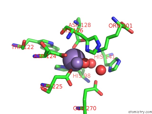
Manganese binding site 1 out of 4 in 5zeh
Go back to
Manganese binding site 1 out
of 4 in the Crystal Structure of Entamoeba Histolytica Arginase in Complex with L- Ornithine at 2.35 A
Mono view

Stereo pair view
Mono view
Stereo pair view
A full contact list of Manganese with other atoms in the Mn binding
site number 1 of Crystal Structure of Entamoeba Histolytica Arginase in Complex with L- Ornithine at 2.35 A within 5.0Å range:
|
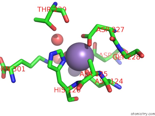
Manganese binding site 2 out of 4 in 5zeh
Go back to
Manganese binding site 2 out
of 4 in the Crystal Structure of Entamoeba Histolytica Arginase in Complex with L- Ornithine at 2.35 A
Mono view

Stereo pair view
Mono view
Stereo pair view
A full contact list of Manganese with other atoms in the Mn binding
site number 2 of Crystal Structure of Entamoeba Histolytica Arginase in Complex with L- Ornithine at 2.35 A within 5.0Å range:
|
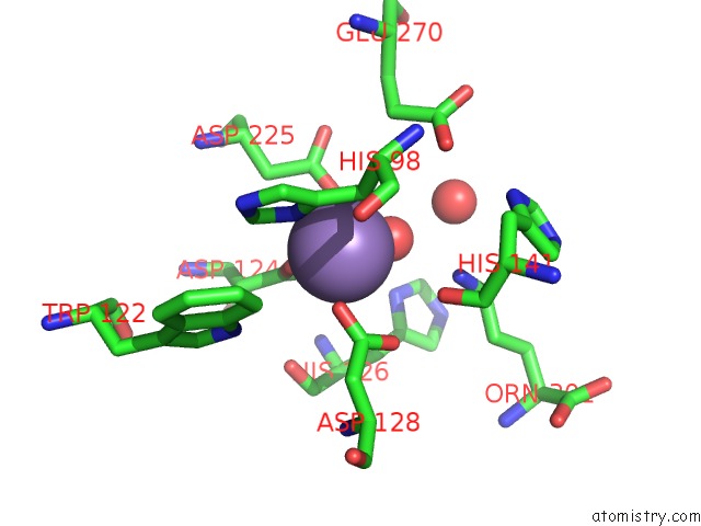
Manganese binding site 3 out of 4 in 5zeh
Go back to
Manganese binding site 3 out
of 4 in the Crystal Structure of Entamoeba Histolytica Arginase in Complex with L- Ornithine at 2.35 A
Mono view

Stereo pair view
Mono view
Stereo pair view
A full contact list of Manganese with other atoms in the Mn binding
site number 3 of Crystal Structure of Entamoeba Histolytica Arginase in Complex with L- Ornithine at 2.35 A within 5.0Å range:
|
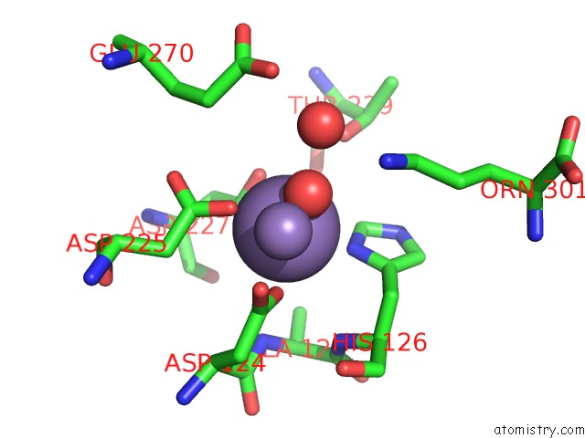
Manganese binding site 4 out of 4 in 5zeh
Go back to
Manganese binding site 4 out
of 4 in the Crystal Structure of Entamoeba Histolytica Arginase in Complex with L- Ornithine at 2.35 A
Mono view

Stereo pair view
Mono view
Stereo pair view
A full contact list of Manganese with other atoms in the Mn binding
site number 4 of Crystal Structure of Entamoeba Histolytica Arginase in Complex with L- Ornithine at 2.35 A within 5.0Å range:
|
Reference:
A.Malik,
V.Dalal,
S.Ankri,
S.Tomar.
Structural Insights Into Entamoeba Histolytica Arginase and Structure-Based Identification of Novel Non-Amino Acid Based Inhibitors As Potential Antiamoebic Molecules. Febs J. V. 286 4135 2019.
ISSN: ISSN 1742-464X
PubMed: 31199070
DOI: 10.1111/FEBS.14960
Page generated: Sun Oct 6 03:41:02 2024
ISSN: ISSN 1742-464X
PubMed: 31199070
DOI: 10.1111/FEBS.14960
Last articles
Zn in 9J0NZn in 9J0O
Zn in 9J0P
Zn in 9FJX
Zn in 9EKB
Zn in 9C0F
Zn in 9CAH
Zn in 9CH0
Zn in 9CH3
Zn in 9CH1