Manganese »
PDB 5z2k-6a9u »
5zbx »
Manganese in PDB 5zbx: The Crystal Structure of the Nucleosome Containing Histone H3.1 Catd(V76Q, K77D)
Protein crystallography data
The structure of The Crystal Structure of the Nucleosome Containing Histone H3.1 Catd(V76Q, K77D), PDB code: 5zbx
was solved by
Y.Arimura,
H.Takagi,
H.Kurumizaka,
with X-Ray Crystallography technique. A brief refinement statistics is given in the table below:
| Resolution Low / High (Å) | 49.28 / 2.58 |
| Space group | P 21 21 21 |
| Cell size a, b, c (Å), α, β, γ (°) | 99.543, 109.003, 170.136, 90.00, 90.00, 90.00 |
| R / Rfree (%) | 21.3 / 24.9 |
Other elements in 5zbx:
The structure of The Crystal Structure of the Nucleosome Containing Histone H3.1 Catd(V76Q, K77D) also contains other interesting chemical elements:
| Chlorine | (Cl) | 2 atoms |
Manganese Binding Sites:
The binding sites of Manganese atom in the The Crystal Structure of the Nucleosome Containing Histone H3.1 Catd(V76Q, K77D)
(pdb code 5zbx). This binding sites where shown within
5.0 Angstroms radius around Manganese atom.
In total 8 binding sites of Manganese where determined in the The Crystal Structure of the Nucleosome Containing Histone H3.1 Catd(V76Q, K77D), PDB code: 5zbx:
Jump to Manganese binding site number: 1; 2; 3; 4; 5; 6; 7; 8;
In total 8 binding sites of Manganese where determined in the The Crystal Structure of the Nucleosome Containing Histone H3.1 Catd(V76Q, K77D), PDB code: 5zbx:
Jump to Manganese binding site number: 1; 2; 3; 4; 5; 6; 7; 8;
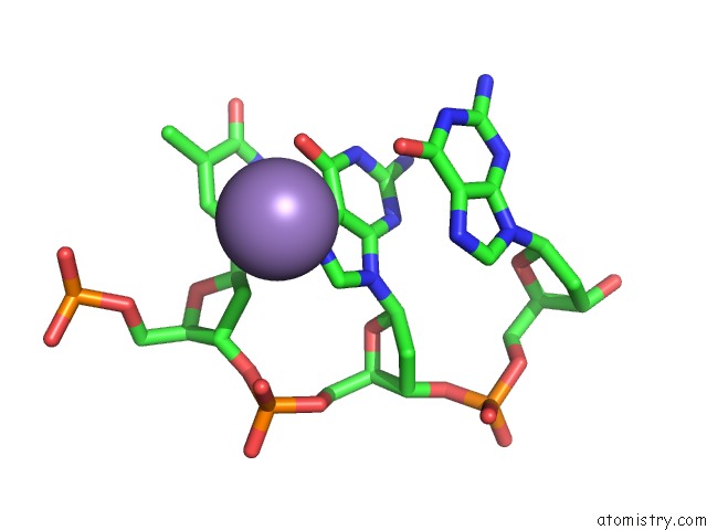
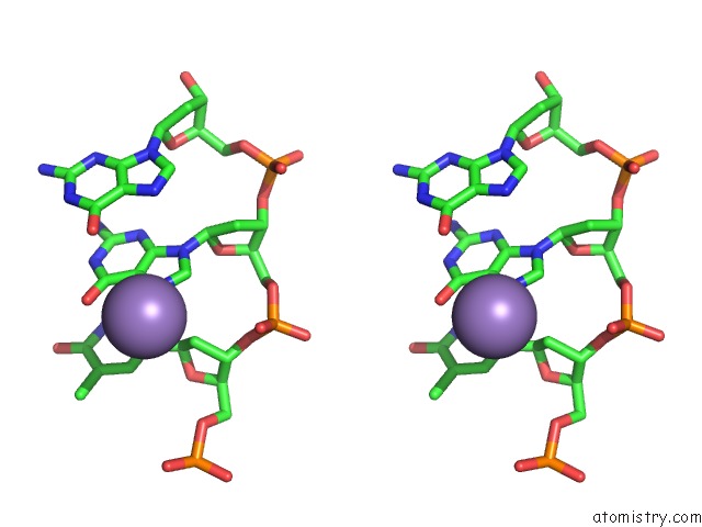
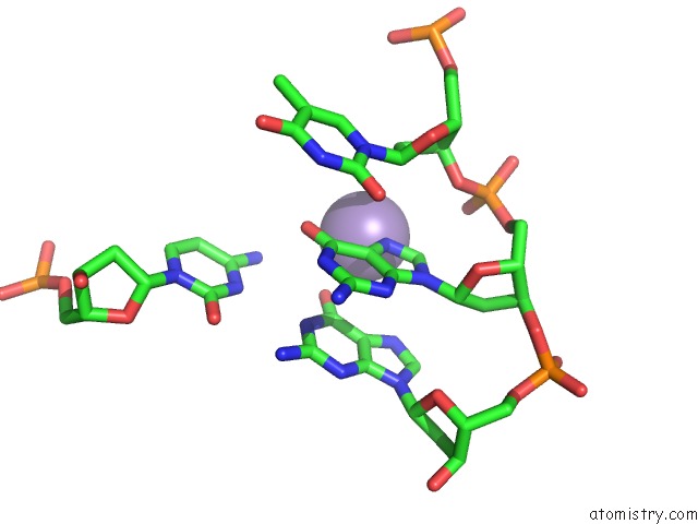
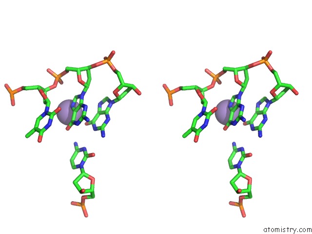
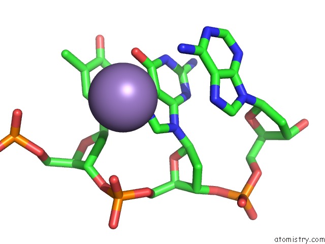
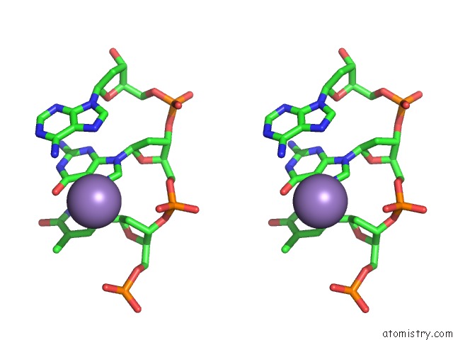
Manganese binding site 1 out of 8 in 5zbx
Go back to
Manganese binding site 1 out
of 8 in the The Crystal Structure of the Nucleosome Containing Histone H3.1 Catd(V76Q, K77D)
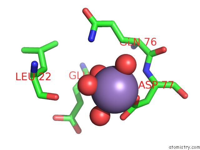
Mono view
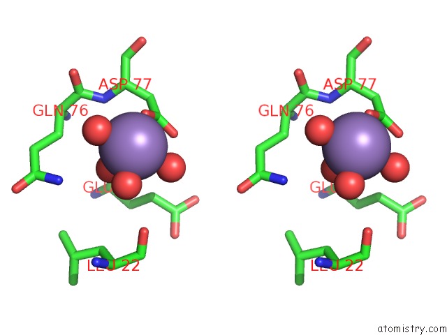
Stereo pair view
Mono view
Stereo pair view
A full contact list of Manganese with other atoms in the Mn binding
site number 1 of The Crystal Structure of the Nucleosome Containing Histone H3.1 Catd(V76Q, K77D) within 5.0Å range:
|
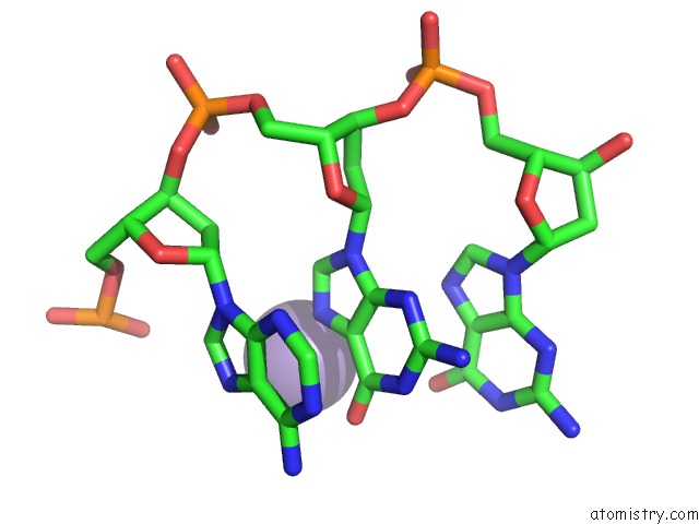
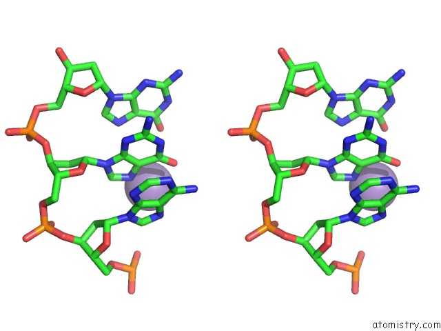
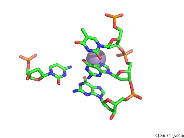
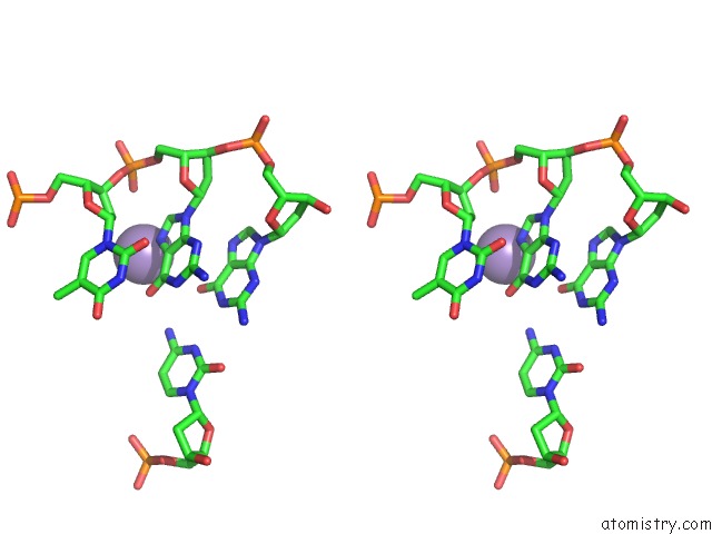
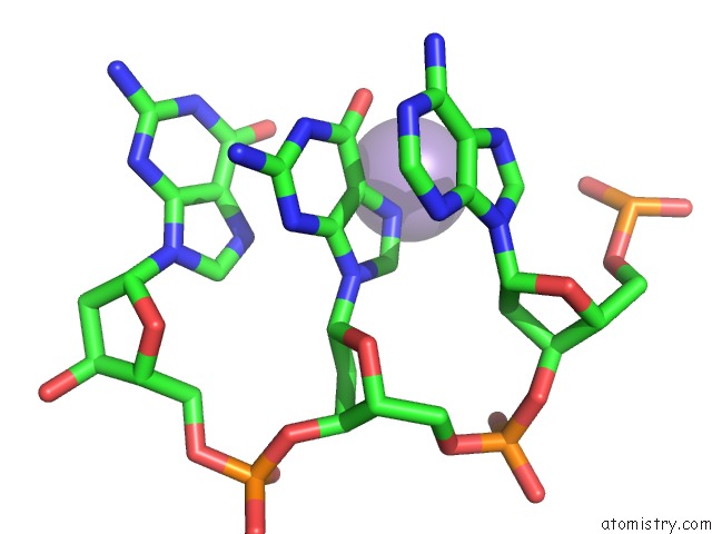
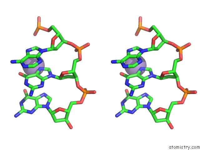
Manganese binding site 2 out of 8 in 5zbx
Go back to
Manganese binding site 2 out
of 8 in the The Crystal Structure of the Nucleosome Containing Histone H3.1 Catd(V76Q, K77D)
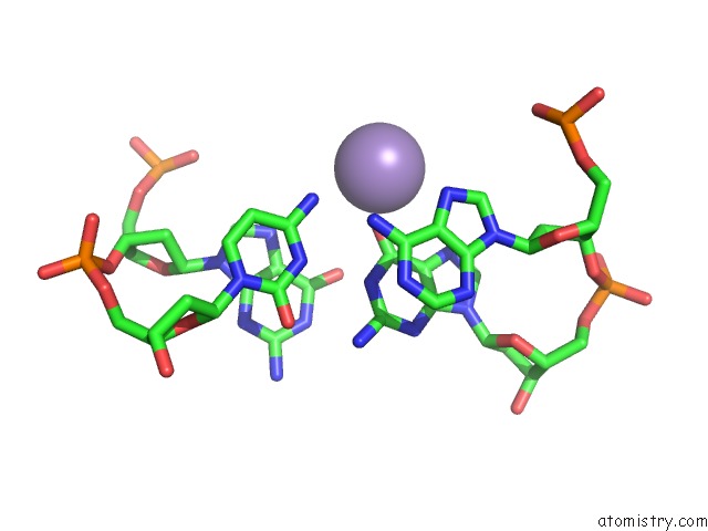
Mono view
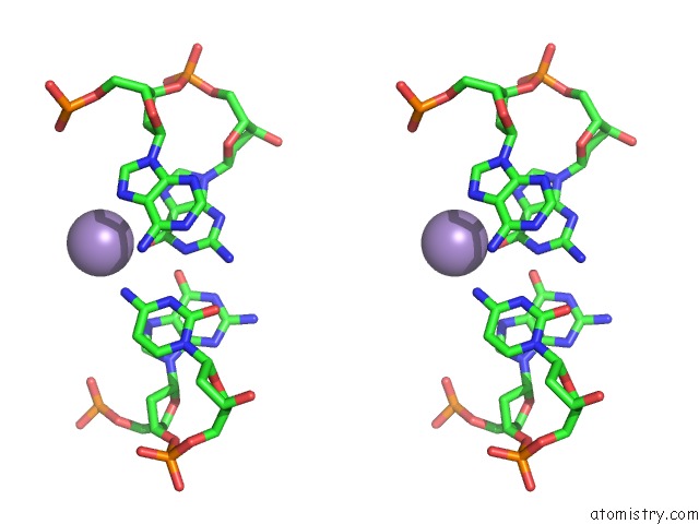
Stereo pair view
Mono view
Stereo pair view
A full contact list of Manganese with other atoms in the Mn binding
site number 2 of The Crystal Structure of the Nucleosome Containing Histone H3.1 Catd(V76Q, K77D) within 5.0Å range:
|
Manganese binding site 3 out of 8 in 5zbx
Go back to
Manganese binding site 3 out
of 8 in the The Crystal Structure of the Nucleosome Containing Histone H3.1 Catd(V76Q, K77D)
Mono view
Stereo pair view
Mono view
Stereo pair view
A full contact list of Manganese with other atoms in the Mn binding
site number 3 of The Crystal Structure of the Nucleosome Containing Histone H3.1 Catd(V76Q, K77D) within 5.0Å range:
|
Manganese binding site 4 out of 8 in 5zbx
Go back to
Manganese binding site 4 out
of 8 in the The Crystal Structure of the Nucleosome Containing Histone H3.1 Catd(V76Q, K77D)
Mono view
Stereo pair view
Mono view
Stereo pair view
A full contact list of Manganese with other atoms in the Mn binding
site number 4 of The Crystal Structure of the Nucleosome Containing Histone H3.1 Catd(V76Q, K77D) within 5.0Å range:
|
Manganese binding site 5 out of 8 in 5zbx
Go back to
Manganese binding site 5 out
of 8 in the The Crystal Structure of the Nucleosome Containing Histone H3.1 Catd(V76Q, K77D)
Mono view
Stereo pair view
Mono view
Stereo pair view
A full contact list of Manganese with other atoms in the Mn binding
site number 5 of The Crystal Structure of the Nucleosome Containing Histone H3.1 Catd(V76Q, K77D) within 5.0Å range:
|
Manganese binding site 6 out of 8 in 5zbx
Go back to
Manganese binding site 6 out
of 8 in the The Crystal Structure of the Nucleosome Containing Histone H3.1 Catd(V76Q, K77D)
Mono view
Stereo pair view
Mono view
Stereo pair view
A full contact list of Manganese with other atoms in the Mn binding
site number 6 of The Crystal Structure of the Nucleosome Containing Histone H3.1 Catd(V76Q, K77D) within 5.0Å range:
|
Manganese binding site 7 out of 8 in 5zbx
Go back to
Manganese binding site 7 out
of 8 in the The Crystal Structure of the Nucleosome Containing Histone H3.1 Catd(V76Q, K77D)
Mono view
Stereo pair view
Mono view
Stereo pair view
A full contact list of Manganese with other atoms in the Mn binding
site number 7 of The Crystal Structure of the Nucleosome Containing Histone H3.1 Catd(V76Q, K77D) within 5.0Å range:
|
Manganese binding site 8 out of 8 in 5zbx
Go back to
Manganese binding site 8 out
of 8 in the The Crystal Structure of the Nucleosome Containing Histone H3.1 Catd(V76Q, K77D)
Mono view
Stereo pair view
Mono view
Stereo pair view
A full contact list of Manganese with other atoms in the Mn binding
site number 8 of The Crystal Structure of the Nucleosome Containing Histone H3.1 Catd(V76Q, K77D) within 5.0Å range:
|
Reference:
Y.Arimura,
H.Tachiwana,
H.Takagi,
T.Hori,
H.Kimura,
T.Fukagawa,
H.Kurumizaka.
The Cenp-A Centromere Targeting Domain Facilitates H4K20 Monomethylation in the Nucleosome By Structural Polymorphism. Nat Commun V. 10 576 2019.
ISSN: ESSN 2041-1723
PubMed: 30718488
DOI: 10.1038/S41467-019-08314-X
Page generated: Sun Oct 6 03:38:47 2024
ISSN: ESSN 2041-1723
PubMed: 30718488
DOI: 10.1038/S41467-019-08314-X
Last articles
Cl in 5WGICl in 5WGV
Cl in 5WGU
Cl in 5WGS
Cl in 5WGT
Cl in 5WGR
Cl in 5WG9
Cl in 5WFZ
Cl in 5WFQ
Cl in 5WFW