Manganese »
PDB 5z2k-6a9u »
5z6w »
Manganese in PDB 5z6w: Crystal Structure of PAFAN1 Bound to 2NT 5'Flap Dna with Gap with Manganese
Enzymatic activity of Crystal Structure of PAFAN1 Bound to 2NT 5'Flap Dna with Gap with Manganese
All present enzymatic activity of Crystal Structure of PAFAN1 Bound to 2NT 5'Flap Dna with Gap with Manganese:
3.1.4.1;
3.1.4.1;
Protein crystallography data
The structure of Crystal Structure of PAFAN1 Bound to 2NT 5'Flap Dna with Gap with Manganese, PDB code: 5z6w
was solved by
H.Jin,
Y.Cho,
with X-Ray Crystallography technique. A brief refinement statistics is given in the table below:
| Resolution Low / High (Å) | 46.99 / 3.20 |
| Space group | P 21 21 21 |
| Cell size a, b, c (Å), α, β, γ (°) | 80.874, 104.969, 105.490, 90.00, 90.00, 90.00 |
| R / Rfree (%) | 18.2 / 23.8 |
Manganese Binding Sites:
The binding sites of Manganese atom in the Crystal Structure of PAFAN1 Bound to 2NT 5'Flap Dna with Gap with Manganese
(pdb code 5z6w). This binding sites where shown within
5.0 Angstroms radius around Manganese atom.
In total 2 binding sites of Manganese where determined in the Crystal Structure of PAFAN1 Bound to 2NT 5'Flap Dna with Gap with Manganese, PDB code: 5z6w:
Jump to Manganese binding site number: 1; 2;
In total 2 binding sites of Manganese where determined in the Crystal Structure of PAFAN1 Bound to 2NT 5'Flap Dna with Gap with Manganese, PDB code: 5z6w:
Jump to Manganese binding site number: 1; 2;
Manganese binding site 1 out of 2 in 5z6w
Go back to
Manganese binding site 1 out
of 2 in the Crystal Structure of PAFAN1 Bound to 2NT 5'Flap Dna with Gap with Manganese
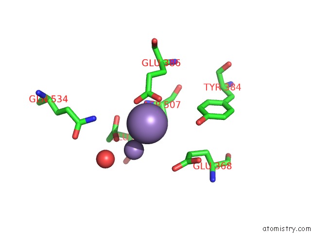
Mono view
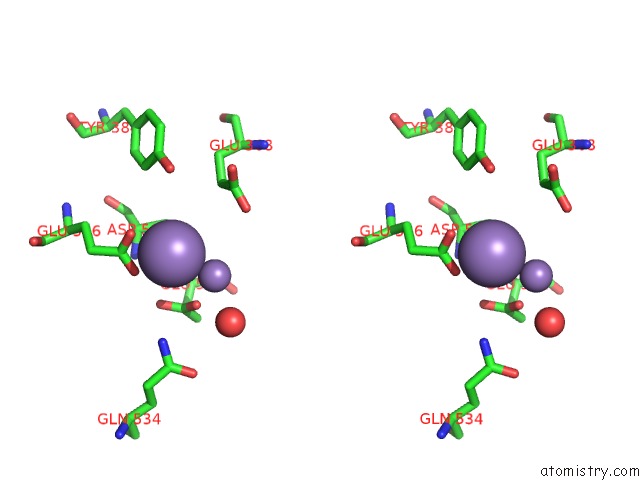
Stereo pair view
Mono view
Stereo pair view
A full contact list of Manganese with other atoms in the Mn binding
site number 1 of Crystal Structure of PAFAN1 Bound to 2NT 5'Flap Dna with Gap with Manganese within 5.0Å range:
|
Manganese binding site 2 out of 2 in 5z6w
Go back to
Manganese binding site 2 out
of 2 in the Crystal Structure of PAFAN1 Bound to 2NT 5'Flap Dna with Gap with Manganese
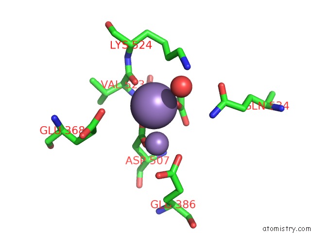
Mono view
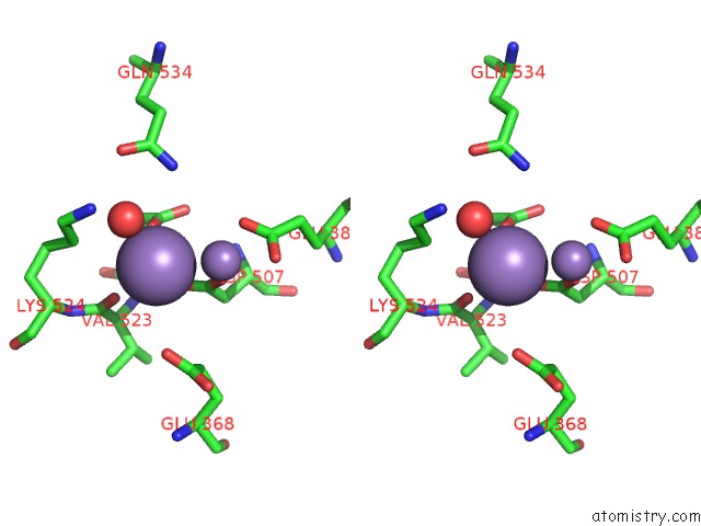
Stereo pair view
Mono view
Stereo pair view
A full contact list of Manganese with other atoms in the Mn binding
site number 2 of Crystal Structure of PAFAN1 Bound to 2NT 5'Flap Dna with Gap with Manganese within 5.0Å range:
|
Reference:
H.Jin,
U.Roy,
G.Lee,
O.D.Scharer,
Y.Cho.
Structural Mechanism of Dna Interstrand Cross-Link Unhooking By the Bacterial FAN1 Nuclease. J. Biol. Chem. V. 293 6482 2018.
ISSN: ESSN 1083-351X
PubMed: 29514982
DOI: 10.1074/JBC.RA118.002171
Page generated: Sun Oct 6 03:38:09 2024
ISSN: ESSN 1083-351X
PubMed: 29514982
DOI: 10.1074/JBC.RA118.002171
Last articles
Zn in 9MJ5Zn in 9HNW
Zn in 9G0L
Zn in 9FNE
Zn in 9DZN
Zn in 9E0I
Zn in 9D32
Zn in 9DAK
Zn in 8ZXC
Zn in 8ZUF