Manganese »
PDB 5wly-5yvs »
5yty »
Manganese in PDB 5yty: Crystal Structure of Echinomycin-D(Acgacgt/Acgtcgt) Complex
Protein crystallography data
The structure of Crystal Structure of Echinomycin-D(Acgacgt/Acgtcgt) Complex, PDB code: 5yty
was solved by
M.H.Hou,
P.C.Wu,
Y.F.Kao,
with X-Ray Crystallography technique. A brief refinement statistics is given in the table below:
| Resolution Low / High (Å) | 20.60 / 1.58 |
| Space group | P 31 1 2 |
| Cell size a, b, c (Å), α, β, γ (°) | 46.362, 46.362, 48.005, 90.00, 90.00, 120.00 |
| R / Rfree (%) | 22.2 / 26.3 |
Other elements in 5yty:
The structure of Crystal Structure of Echinomycin-D(Acgacgt/Acgtcgt) Complex also contains other interesting chemical elements:
| Magnesium | (Mg) | 2 atoms |
Manganese Binding Sites:
The binding sites of Manganese atom in the Crystal Structure of Echinomycin-D(Acgacgt/Acgtcgt) Complex
(pdb code 5yty). This binding sites where shown within
5.0 Angstroms radius around Manganese atom.
In total only one binding site of Manganese was determined in the Crystal Structure of Echinomycin-D(Acgacgt/Acgtcgt) Complex, PDB code: 5yty:
In total only one binding site of Manganese was determined in the Crystal Structure of Echinomycin-D(Acgacgt/Acgtcgt) Complex, PDB code: 5yty:
Manganese binding site 1 out of 1 in 5yty
Go back to
Manganese binding site 1 out
of 1 in the Crystal Structure of Echinomycin-D(Acgacgt/Acgtcgt) Complex
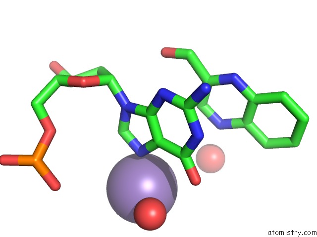
Mono view
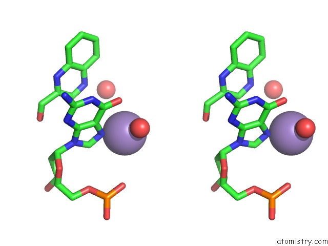
Stereo pair view
Mono view
Stereo pair view
A full contact list of Manganese with other atoms in the Mn binding
site number 1 of Crystal Structure of Echinomycin-D(Acgacgt/Acgtcgt) Complex within 5.0Å range:
|
Reference:
P.C.Wu,
S.L.Tzeng,
C.K.Chang,
Y.F.Kao,
M.J.Waring,
M.H.Hou.
Cooperative Recognition of T:T Mismatch By Echinomycin Causes Structural Distortions in Dna Duplex Nucleic Acids Res. V. 46 7396 2018.
ISSN: ESSN 1362-4962
PubMed: 29741655
DOI: 10.1093/NAR/GKY345
Page generated: Sun Oct 6 03:33:20 2024
ISSN: ESSN 1362-4962
PubMed: 29741655
DOI: 10.1093/NAR/GKY345
Last articles
Zn in 9J0NZn in 9J0O
Zn in 9J0P
Zn in 9FJX
Zn in 9EKB
Zn in 9C0F
Zn in 9CAH
Zn in 9CH0
Zn in 9CH3
Zn in 9CH1