Manganese »
PDB 5wly-5yvs »
5x2j »
Manganese in PDB 5x2j: Crystal Structure of A Recombinant Hybrid Manganese Superoxide Dismutase From Staphylococcus Equorum and Staphylococcus Saprophyticus
Protein crystallography data
The structure of Crystal Structure of A Recombinant Hybrid Manganese Superoxide Dismutase From Staphylococcus Equorum and Staphylococcus Saprophyticus, PDB code: 5x2j
was solved by
D.S.Retnoningrum,
H.Yoshida,
S.Arumsari,
S.Kamitori,
W.T.Ismaya,
with X-Ray Crystallography technique. A brief refinement statistics is given in the table below:
| Resolution Low / High (Å) | 36.21 / 1.40 |
| Space group | P 32 2 1 |
| Cell size a, b, c (Å), α, β, γ (°) | 57.362, 57.362, 105.762, 90.00, 90.00, 120.00 |
| R / Rfree (%) | 14.2 / 18.5 |
Manganese Binding Sites:
The binding sites of Manganese atom in the Crystal Structure of A Recombinant Hybrid Manganese Superoxide Dismutase From Staphylococcus Equorum and Staphylococcus Saprophyticus
(pdb code 5x2j). This binding sites where shown within
5.0 Angstroms radius around Manganese atom.
In total only one binding site of Manganese was determined in the Crystal Structure of A Recombinant Hybrid Manganese Superoxide Dismutase From Staphylococcus Equorum and Staphylococcus Saprophyticus, PDB code: 5x2j:
In total only one binding site of Manganese was determined in the Crystal Structure of A Recombinant Hybrid Manganese Superoxide Dismutase From Staphylococcus Equorum and Staphylococcus Saprophyticus, PDB code: 5x2j:
Manganese binding site 1 out of 1 in 5x2j
Go back to
Manganese binding site 1 out
of 1 in the Crystal Structure of A Recombinant Hybrid Manganese Superoxide Dismutase From Staphylococcus Equorum and Staphylococcus Saprophyticus
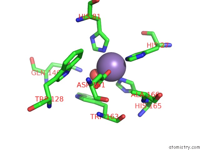
Mono view
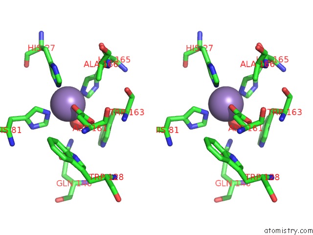
Stereo pair view
Mono view
Stereo pair view
A full contact list of Manganese with other atoms in the Mn binding
site number 1 of Crystal Structure of A Recombinant Hybrid Manganese Superoxide Dismutase From Staphylococcus Equorum and Staphylococcus Saprophyticus within 5.0Å range:
|
Reference:
D.S.Retnoningrum,
H.Yoshida,
S.Arumsari,
S.Kamitori,
W.T.Ismaya.
The First Crystal Structure of Manganese Superoxide Dismutase From the Genus Staphylococcus Acta Crystallogr F Struct V. 74 135 2018BIOL Commun.
ISSN: ESSN 2053-230X
PubMed: 29497016
DOI: 10.1107/S2053230X18001036
Page generated: Sun Oct 6 03:23:27 2024
ISSN: ESSN 2053-230X
PubMed: 29497016
DOI: 10.1107/S2053230X18001036
Last articles
Zn in 9J0NZn in 9J0O
Zn in 9J0P
Zn in 9FJX
Zn in 9EKB
Zn in 9C0F
Zn in 9CAH
Zn in 9CH0
Zn in 9CH3
Zn in 9CH1