Manganese »
PDB 5vqn-5wg9 »
5w44 »
Manganese in PDB 5w44: Crystal Structure of the Influenza Virus Pa Endonuclease in Complex with Inhibitor 7A (Sri-29770)
Protein crystallography data
The structure of Crystal Structure of the Influenza Virus Pa Endonuclease in Complex with Inhibitor 7A (Sri-29770), PDB code: 5w44
was solved by
G.Kumar,
S.W.White,
with X-Ray Crystallography technique. A brief refinement statistics is given in the table below:
| Resolution Low / High (Å) | 35.59 / 2.10 |
| Space group | P 64 2 2 |
| Cell size a, b, c (Å), α, β, γ (°) | 74.111, 74.111, 127.845, 90.00, 90.00, 120.00 |
| R / Rfree (%) | 19.3 / 22.4 |
Other elements in 5w44:
The structure of Crystal Structure of the Influenza Virus Pa Endonuclease in Complex with Inhibitor 7A (Sri-29770) also contains other interesting chemical elements:
| Magnesium | (Mg) | 1 atom |
| Chlorine | (Cl) | 2 atoms |
| Sodium | (Na) | 1 atom |
Manganese Binding Sites:
The binding sites of Manganese atom in the Crystal Structure of the Influenza Virus Pa Endonuclease in Complex with Inhibitor 7A (Sri-29770)
(pdb code 5w44). This binding sites where shown within
5.0 Angstroms radius around Manganese atom.
In total only one binding site of Manganese was determined in the Crystal Structure of the Influenza Virus Pa Endonuclease in Complex with Inhibitor 7A (Sri-29770), PDB code: 5w44:
In total only one binding site of Manganese was determined in the Crystal Structure of the Influenza Virus Pa Endonuclease in Complex with Inhibitor 7A (Sri-29770), PDB code: 5w44:
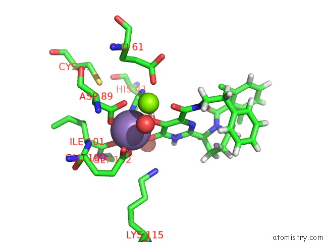
Manganese binding site 1 out of 1 in 5w44
Go back to
Manganese binding site 1 out
of 1 in the Crystal Structure of the Influenza Virus Pa Endonuclease in Complex with Inhibitor 7A (Sri-29770)
Mono view

Stereo pair view
Mono view
Stereo pair view
A full contact list of Manganese with other atoms in the Mn binding
site number 1 of Crystal Structure of the Influenza Virus Pa Endonuclease in Complex with Inhibitor 7A (Sri-29770) within 5.0Å range:
|
Reference:
D.Beylkin,
G.Kumar,
W.Zhou,
J.Park,
T.Jeevan,
C.Lagisetti,
R.Harfoot,
R.J.Webby,
S.W.White,
T.R.Webb.
Protein-Structure Assisted Optimization of 4,5-Dihydroxypyrimidine-6-Carboxamide Inhibitors of Influenza Virus Endonuclease. Sci Rep V. 7 17139 2017.
ISSN: ESSN 2045-2322
PubMed: 29215062
DOI: 10.1038/S41598-017-17419-6
Page generated: Sun Oct 6 03:14:17 2024
ISSN: ESSN 2045-2322
PubMed: 29215062
DOI: 10.1038/S41598-017-17419-6
Last articles
Zn in 9J0NZn in 9J0O
Zn in 9J0P
Zn in 9FJX
Zn in 9EKB
Zn in 9C0F
Zn in 9CAH
Zn in 9CH0
Zn in 9CH3
Zn in 9CH1