Manganese »
PDB 5uqt-5vpx »
5v07 »
Manganese in PDB 5v07: Crystal Structure of Human Exonuclease 1 EXO1 (D173A) in Complex with 5' Recessed-End Dna (Rv)
Protein crystallography data
The structure of Crystal Structure of Human Exonuclease 1 EXO1 (D173A) in Complex with 5' Recessed-End Dna (Rv), PDB code: 5v07
was solved by
Y.Shi,
L.S.Beese,
with X-Ray Crystallography technique. A brief refinement statistics is given in the table below:
| Resolution Low / High (Å) | 36.92 / 2.15 |
| Space group | P 43 21 2 |
| Cell size a, b, c (Å), α, β, γ (°) | 73.837, 73.837, 179.067, 90.00, 90.00, 90.00 |
| R / Rfree (%) | 17.6 / 22 |
Other elements in 5v07:
The structure of Crystal Structure of Human Exonuclease 1 EXO1 (D173A) in Complex with 5' Recessed-End Dna (Rv) also contains other interesting chemical elements:
| Sodium | (Na) | 1 atom |
Manganese Binding Sites:
The binding sites of Manganese atom in the Crystal Structure of Human Exonuclease 1 EXO1 (D173A) in Complex with 5' Recessed-End Dna (Rv)
(pdb code 5v07). This binding sites where shown within
5.0 Angstroms radius around Manganese atom.
In total 4 binding sites of Manganese where determined in the Crystal Structure of Human Exonuclease 1 EXO1 (D173A) in Complex with 5' Recessed-End Dna (Rv), PDB code: 5v07:
Jump to Manganese binding site number: 1; 2; 3; 4;
In total 4 binding sites of Manganese where determined in the Crystal Structure of Human Exonuclease 1 EXO1 (D173A) in Complex with 5' Recessed-End Dna (Rv), PDB code: 5v07:
Jump to Manganese binding site number: 1; 2; 3; 4;
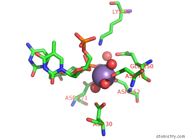
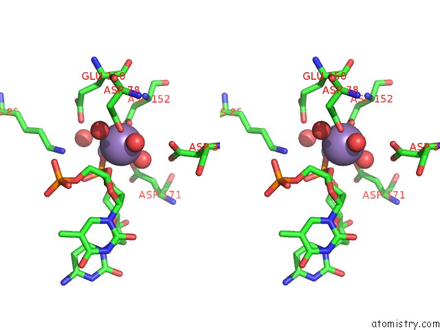
Manganese binding site 1 out of 4 in 5v07
Go back to
Manganese binding site 1 out
of 4 in the Crystal Structure of Human Exonuclease 1 EXO1 (D173A) in Complex with 5' Recessed-End Dna (Rv)
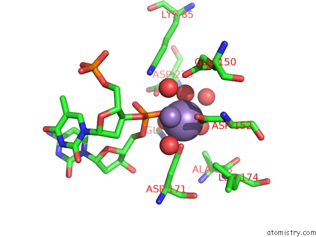
Mono view
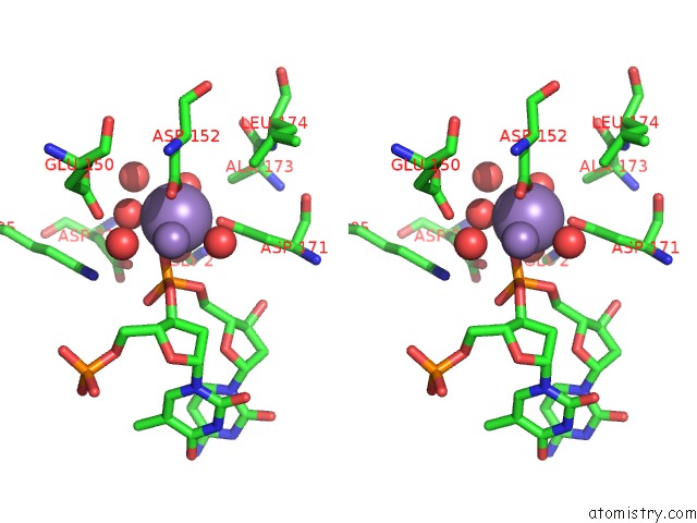
Stereo pair view
Mono view
Stereo pair view
A full contact list of Manganese with other atoms in the Mn binding
site number 1 of Crystal Structure of Human Exonuclease 1 EXO1 (D173A) in Complex with 5' Recessed-End Dna (Rv) within 5.0Å range:
|
Manganese binding site 2 out of 4 in 5v07
Go back to
Manganese binding site 2 out
of 4 in the Crystal Structure of Human Exonuclease 1 EXO1 (D173A) in Complex with 5' Recessed-End Dna (Rv)
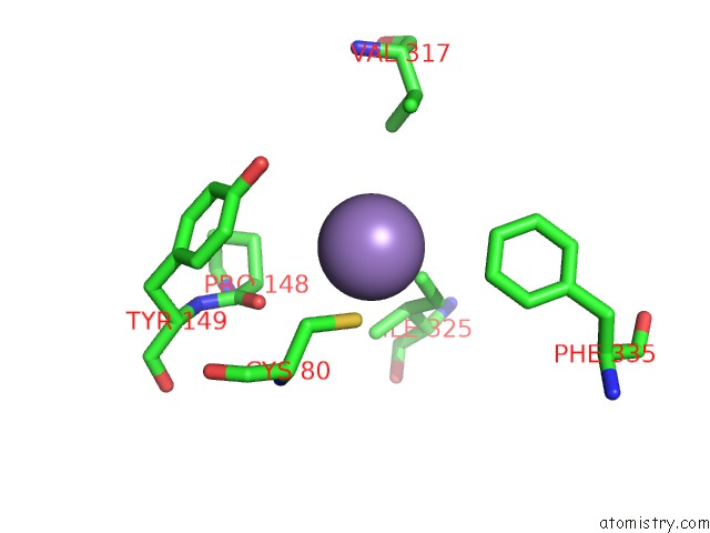
Mono view
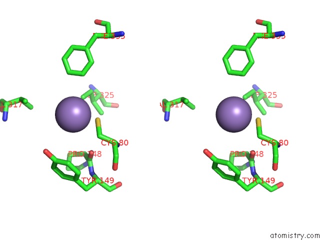
Stereo pair view
Mono view
Stereo pair view
A full contact list of Manganese with other atoms in the Mn binding
site number 2 of Crystal Structure of Human Exonuclease 1 EXO1 (D173A) in Complex with 5' Recessed-End Dna (Rv) within 5.0Å range:
|
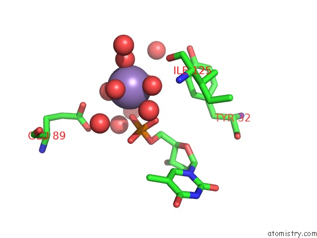
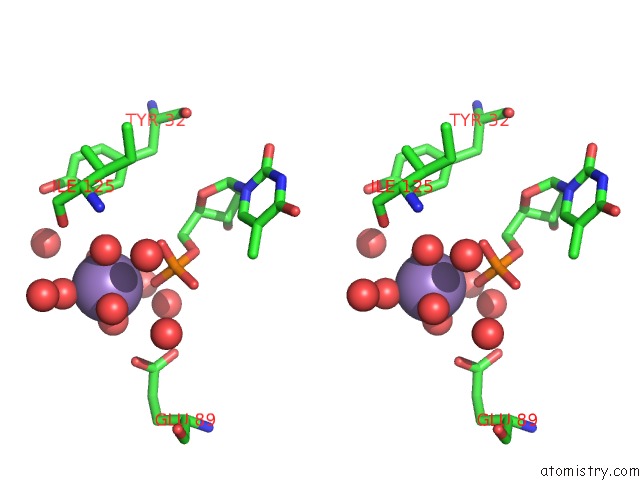
Manganese binding site 3 out of 4 in 5v07
Go back to
Manganese binding site 3 out
of 4 in the Crystal Structure of Human Exonuclease 1 EXO1 (D173A) in Complex with 5' Recessed-End Dna (Rv)
Mono view
Stereo pair view
Mono view
Stereo pair view
A full contact list of Manganese with other atoms in the Mn binding
site number 3 of Crystal Structure of Human Exonuclease 1 EXO1 (D173A) in Complex with 5' Recessed-End Dna (Rv) within 5.0Å range:
|
Manganese binding site 4 out of 4 in 5v07
Go back to
Manganese binding site 4 out
of 4 in the Crystal Structure of Human Exonuclease 1 EXO1 (D173A) in Complex with 5' Recessed-End Dna (Rv)
Mono view
Stereo pair view
Mono view
Stereo pair view
A full contact list of Manganese with other atoms in the Mn binding
site number 4 of Crystal Structure of Human Exonuclease 1 EXO1 (D173A) in Complex with 5' Recessed-End Dna (Rv) within 5.0Å range:
|
Reference:
Y.Shi,
H.W.Hellinga,
L.S.Beese.
Interplay of Catalysis, Fidelity, Threading, and Processivity in the Exo- and Endonucleolytic Reactions of Human Exonuclease I. Proc. Natl. Acad. Sci. V. 114 6010 2017U.S.A..
ISSN: ESSN 1091-6490
PubMed: 28533382
DOI: 10.1073/PNAS.1704845114
Page generated: Sun Oct 6 03:06:31 2024
ISSN: ESSN 1091-6490
PubMed: 28533382
DOI: 10.1073/PNAS.1704845114
Last articles
Fe in 2YXOFe in 2YRS
Fe in 2YXC
Fe in 2YNM
Fe in 2YVJ
Fe in 2YP1
Fe in 2YU2
Fe in 2YU1
Fe in 2YQB
Fe in 2YOO