Manganese »
PDB 5svc-5uqn »
5twq »
Manganese in PDB 5twq: Post-Catalytic Nicked Complex of Human Polymerase Mu with Newly Incorporated Utp
Protein crystallography data
The structure of Post-Catalytic Nicked Complex of Human Polymerase Mu with Newly Incorporated Utp, PDB code: 5twq
was solved by
A.F.Moon,
J.M.Pryor,
D.A.Ramsden,
T.A.Kunkel,
K.Bebenek,
L.C.Pedersen,
with X-Ray Crystallography technique. A brief refinement statistics is given in the table below:
| Resolution Low / High (Å) | 34.97 / 1.80 |
| Space group | P 21 21 21 |
| Cell size a, b, c (Å), α, β, γ (°) | 59.969, 68.712, 110.413, 90.00, 90.00, 90.00 |
| R / Rfree (%) | 16.8 / 20.5 |
Other elements in 5twq:
The structure of Post-Catalytic Nicked Complex of Human Polymerase Mu with Newly Incorporated Utp also contains other interesting chemical elements:
| Magnesium | (Mg) | 1 atom |
| Chlorine | (Cl) | 1 atom |
| Sodium | (Na) | 1 atom |
Manganese Binding Sites:
The binding sites of Manganese atom in the Post-Catalytic Nicked Complex of Human Polymerase Mu with Newly Incorporated Utp
(pdb code 5twq). This binding sites where shown within
5.0 Angstroms radius around Manganese atom.
In total only one binding site of Manganese was determined in the Post-Catalytic Nicked Complex of Human Polymerase Mu with Newly Incorporated Utp, PDB code: 5twq:
In total only one binding site of Manganese was determined in the Post-Catalytic Nicked Complex of Human Polymerase Mu with Newly Incorporated Utp, PDB code: 5twq:
Manganese binding site 1 out of 1 in 5twq
Go back to
Manganese binding site 1 out
of 1 in the Post-Catalytic Nicked Complex of Human Polymerase Mu with Newly Incorporated Utp
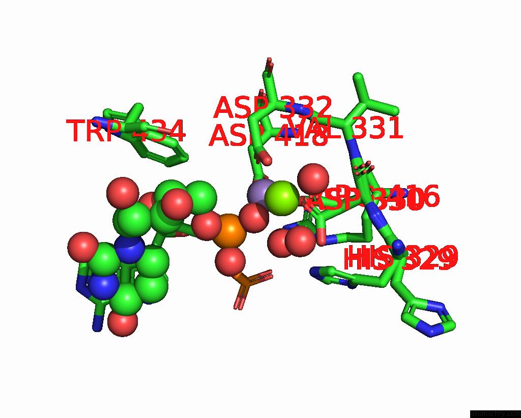
Mono view
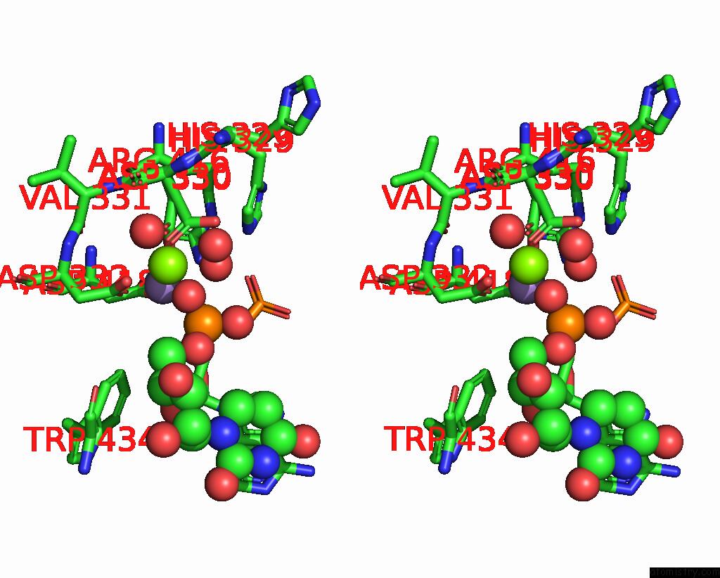
Stereo pair view
Mono view
Stereo pair view
A full contact list of Manganese with other atoms in the Mn binding
site number 1 of Post-Catalytic Nicked Complex of Human Polymerase Mu with Newly Incorporated Utp within 5.0Å range:
|
Reference:
A.F.Moon,
J.M.Pryor,
D.A.Ramsden,
T.A.Kunkel,
K.Bebenek,
L.C.Pedersen.
Structural Accommodation of Ribonucleotide Incorporation By the Dna Repair Enzyme Polymerase Mu. Nucleic Acids Res. V. 45 9138 2017.
ISSN: ESSN 1362-4962
PubMed: 28911097
DOI: 10.1093/NAR/GKX527
Page generated: Sat Aug 16 19:14:49 2025
ISSN: ESSN 1362-4962
PubMed: 28911097
DOI: 10.1093/NAR/GKX527
Last articles
Mo in 8BTSMo in 8BQR
Mo in 8BQQ
Mo in 8BQP
Mo in 7Z5J
Mo in 7ZUB
Mo in 7Z0T
Mo in 7WY3
Mo in 7WY2
Mo in 7WY1